Application
Immunoperoxidase staining of formalin fixed, paraffin-embedded human urinary bladder tissue showing cytoplasmic and membrane staining of urothelial cells.Application
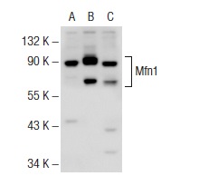
Western blot analysis of Mfn1 expression in non-transfected 293T: (A), human Mfn1 transfected 293T: (B) and HeLa (C) whole cell lysates.Application
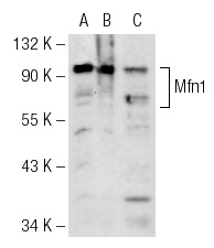
Western blot analysis of Mfn1 expression in non-transfected 293T: (A), mouse Mfn1 transfected 293T: (B) and HeLa (C) whole cell lysates.IF
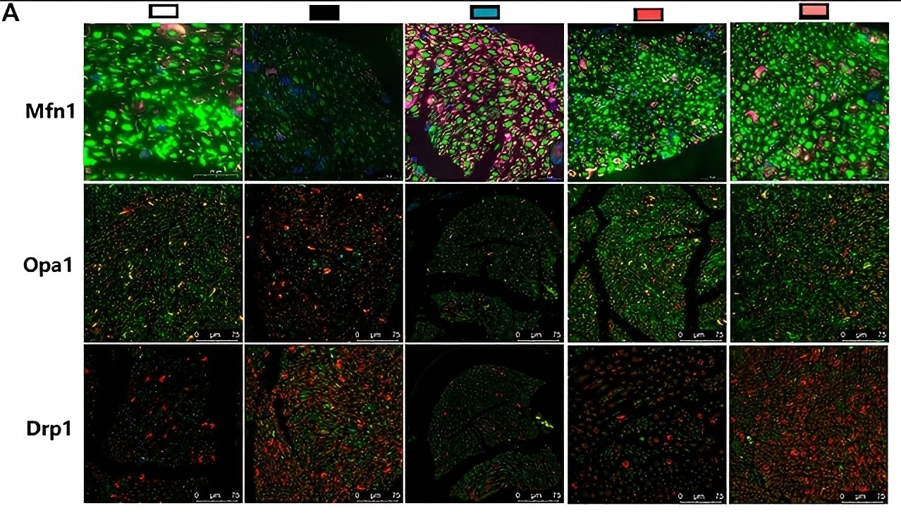
(A) Representative images of immunofluorescence staining on Mfn1 (green); scale bar, 20 μm. Opa1 (green); scale bar, 75 μm. Drp1 (green); scale bar, 75 μm. Schwann cells are stained with S100β (red).WB
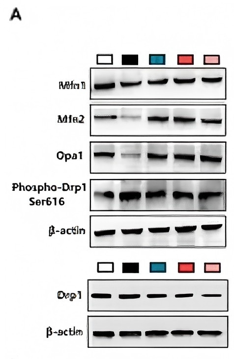
(A)Western blotting of Mfn1, Mfn2, Opa1, Drp1, and phospho-Drp1 (Ser616) of SCs and quantifications of these proteins. The results were normalized to the values of the 25 mM glucose group. β-actin images are reused| Product Name | Mfn1 Antibody (H-65) |
|---|---|
| Antibody Type | Primary Antibodies |
| Modification Notes | Mitofusin 1 (Mfn1) and mitofusin 2 (Mfn2) are homologs for the Drosophila protein fuzzy onion (Fzo). They are mitochondrial membrane proteins and are mediators of mitochondrial fusion. A GTPase domain is required for Mfn protein function but the molecular mechanisms of the GTPase-dependent reaction as well as the functional division of the two Mfn proteins are unknown. They are essential for embryonic development and may play a role in the pathobiology of obesity. Although the Mfn1 and Mfn2 genes are broadly expressed, they show different levels of expression in different tissues. Two Mfn1 transcripts are elevated in heart, while Mfn2 mRNA is abundantly expressed in heart and muscle tissue but present only at low levels in many other tissues. Mfn1 localizes to mitochondria and participates in at least two different high molecular weight protein complexes in a GTP-dependent manner. Purified recombinant Mfn1 exhibited approximately eightfold higher GTPase activity than Mfn2. |
| Clonality | Polyclonal |
|---|---|
| Isotype | IgG |
| Host Species | Rabbit |
| Tested Applications | ELISAIFIHCIPWB |
| recommended for detection of Mitofusin 1 of mouse, rat and human origin by WB, IP, IF, IHC(P) and ELISA also reactive with additional species, including equine, canine and bovine: |
|
| Species Reactivity | HumanMouseRat |
| Concentration | 1mg/ml |
| Purification | Affinity purified |
| Alternative Names | epitope corresponding to amino acids 10-74 mapping within an N-terminal cytoplasmic domain of Mfn1 of human origin |
|---|---|
| Tissue Specificity | epitope corresponding to amino acids 10-74 mapping within an N-terminal cytoplasmic domain of Mfn1 of human origin |
| Entrez Gene | 55669 |
|---|
Application
Immunoperoxidase staining of formalin fixed, paraffin-embedded human urinary bladder tissue showing cytoplasmic and membrane staining of urothelial cells.
Application
Western blot analysis of Mfn1 expression in non-transfected 293T: (A), human Mfn1 transfected 293T: (B) and HeLa (C) whole cell lysates.
Application
Western blot analysis of Mfn1 expression in non-transfected 293T: (A), mouse Mfn1 transfected 293T: (B) and HeLa (C) whole cell lysates.
IF
(A) Representative images of immunofluorescence staining on Mfn1 (green); scale bar, 20 μm. Opa1 (green); scale bar, 75 μm. Drp1 (green); scale bar, 75 μm. Schwann cells are stained with S100β (red).
WB
(A)Western blotting of Mfn1, Mfn2, Opa1, Drp1, and phospho-Drp1 (Ser616) of SCs and quantifications of these proteins. The results were normalized to the values of the 25 mM glucose group. β-actin images are reused| Application Notes | recommended for detection of Mitofusin 1 of mouse, rat and human origin by WB, IP, IF, IHC(P) and ELISA also reactive with additional species, including equine, canine and bovine: |
|---|
| Form | Liquid |
|---|---|
| Storage Instructions | For short-term storage, store at 4° C. For long-term storage, aliquot and store at -20ºC or below. Avoid multiple freeze-thaw cycles. |
| Storage Buffer | phosphate buffered saline , pH 7.4, 150mM NaCl, 0.02% sodium azide and 50% glycerol. |


