Application
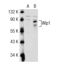
Western blot analysis of Wip1 expression in non-transfected: (A) and human Wip1 transfected: (B) 293T whole cell lysates.Application
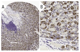
Immunoperoxidase staining of formalin fixed, paraffin-embedded human stomach tissue showing cytoplasmic staining of glandular cells at low (A) and high (B) magnification. Kindly provided by The Swedish Human Protein Atlas (HPA) program.Application
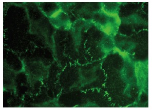
Western blot analysis of Wip1 expression in F9 whole cell lysate| Product Name | Wip1 Antibody (H-300) |
|---|---|
| Antibody Type | Primary Antibodies |
| Modification Notes | Several major serine/threonine protein phosphatases have been identified in eukaryotic cells. These include protein phosphatase families 1, 2A, 2B, 2C, X and Y (PP-1, PP-2A, PP-2B, PP-2C, PP-X and PP-Y). These enzymes can be distinguished by their action on phosphorylase kinase and their sensitivity to certain activators and inhibitors. Wip1 (wildtype p53-induced phosphatase or PPM1D), a protein identified in the p53 DNA response pathway, is a member of the PP-2C family. Wip1 is a serine/threonine protein phosphatase which selectively inactivates p38 MAPK and dephosphorylates the ATM/ATR targets, Chk1 and p53. Wip1 is ubiquitously expressed but is present at very high levels in testis. Deletion of Wip1 results in a reduction of T and B cell function and compromised cell division, rendering cells resistant to becoming cancerous and slowing tumor development. |
| Clonality | Polyclonal |
|---|---|
| Isotype | IgG |
| Host Species | Rabbit |
| Tested Applications | WBIPIFIHCELISA |
| recommended for detection of Wip1 of mouse, rat and human origin by WB, IP, IF, IHC(P) and ELISA also reactive with additional species, including equine, canine, bovine and porcine | |
| Species Reactivity | HumanMouseRat |
| Concentration | 1mg/ml |
| Purification | Affinity purified |
| Alternative Names | epitope corresponding to amino acids 306-605 mapping at the C-terminus of Wip1 of human origin |
|---|---|
| Tissue Specificity | epitope corresponding to amino acids 306-605 mapping at the C-terminus of Wip1 of human origin |
| Entrez Gene | 8493 |
|---|
Application
Western blot analysis of Wip1 expression in non-transfected: (A) and human Wip1 transfected: (B) 293T whole cell lysates.
Application
Immunoperoxidase staining of formalin fixed, paraffin-embedded human stomach tissue showing cytoplasmic staining of glandular cells at low (A) and high (B) magnification. Kindly provided by The Swedish Human Protein Atlas (HPA) program.
Application
Western blot analysis of Wip1 expression in F9 whole cell lysate| Application Notes | recommended for detection of Wip1 of mouse, rat and human origin by WB, IP, IF, IHC(P) and ELISA also reactive with additional species, including equine, canine, bovine and porcine |
|---|
| Form | Liquid |
|---|---|
| Storage Instructions | For short-term storage, store at 4° C. For long-term storage, aliquot and store at -20ºC or below. Avoid multiple freeze-thaw cycles. |
| Storage Buffer | phosphate buffered saline , pH 7.4, 150mM NaCl, 0.02% sodium azide and 50% glycerol. |


