Application
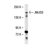
Western blot analysis of JMJD3 expression in 293 whole cell lysates.IF
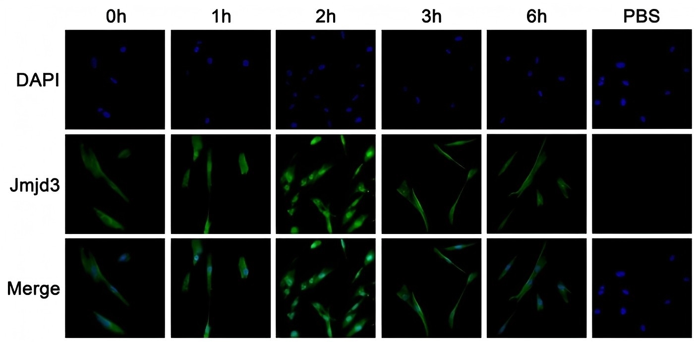
Immunofluorescent staining of Jmjd3 in inflamed HPDLs. HPDLs were treated with lipopolysaccharide (10 ng/mL) at different time points and immunostained with the anti-Jmjd3 antibody. Notably, lipopolysaccharide treatment promoted Jmjd3 nuclear accumulation transiently and reversible. Nuclei were labeled with DAPI (4′,6-diamidino-2-phenylindole) (blue) and Jmjd3 was labeled with fluorescein isothiocyanate (green). (For interpretation of the references to colour in this figure legend, the reader is referred to the web version of this article).IHC
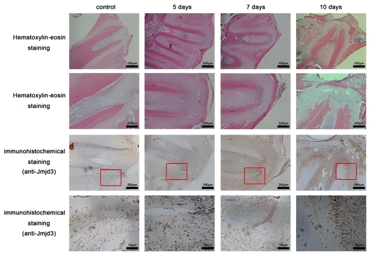
Expression of Jmjd3 in the early stage of apical periodontitis. Hematoxylin-eosin staining showed in the experimental group at day 5, significant inflammatory cell infiltration was observed at the middle part of the pulp tissue. In the group at day 7, slight inflammatory cell infiltration was observed in the periapical tissue. And in the group at day 10, significant inflammatory cell infiltration was observed in the periapical tissue. These results indicated that apical periodontitis was successfully established after the pulp chamber was left open for 7 days and 10 days. Meanwhile, immunohistochemical revealed Jmjd3 was up-regulated significantly in periodontal ligament cells in the group at day 7 and 10.| Product Name | JMJD3 Antibody (N-24) |
|---|---|
| Antibody Type | Primary Antibodies |
| Modification Notes | JMJD3 (jumonji domain containing 3), also known as KDM6B (Lysine demethylase 6B), is a 1,679 amino acid nuclear protein that contains one JmjC domain and belongs to the highly conserved JmjC domain-containing protein family. Functioning as a histone demethylase, JMJD3 uses iron and ascorbate as cofactors to demethylate dimethylated and trimethylated Lys-27 residues of Histone H3, thereby playing an important role in the modification of the histone code. Additionally, JMJD3 regulates posterior development and is involved in the inflammatory response, specifically by mediating macrophage differentiation. JMJD3 is also thought to control the expression of neurogenesis-related proteins and, via this regulatory mechanism, may be necessary for neural commitment during early development. Two isoforms of JMJD3 exist due to alternative splicing events. |
| Clonality | Polyclonal |
|---|---|
| Isotype | IgG |
| Host Species | Rabbit |
| Tested Applications | ELISAIFIHCIPWB |
| WB:1:100~1:500 IP:1:20~1:100 IF:1:20~1:100 IHC:1:10~1:50 |
|
| Species Reactivity | HumanMouseRat |
| Concentration | 1mg/ml |
| Purification | Affinity purified |
| Alternative Names | recommended for detection of JMJD3 of human origin by WB IP IF IHC(P) and ELISA |
|---|---|
| Molecular Weight(MW) | 180kDa |
| Tissue Specificity | recommended for detection of JMJD3 of human origin by WB, IP, IF, IHC(P) and ELISA |
Application
Western blot analysis of JMJD3 expression in 293 whole cell lysates.
IF
Immunofluorescent staining of Jmjd3 in inflamed HPDLs. HPDLs were treated with lipopolysaccharide (10 ng/mL) at different time points and immunostained with the anti-Jmjd3 antibody. Notably, lipopolysaccharide treatment promoted Jmjd3 nuclear accumulation transiently and reversible. Nuclei were labeled with DAPI (4′,6-diamidino-2-phenylindole) (blue) and Jmjd3 was labeled with fluorescein isothiocyanate (green). (For interpretation of the references to colour in this figure legend, the reader is referred to the web version of this article).
IHC
Expression of Jmjd3 in the early stage of apical periodontitis. Hematoxylin-eosin staining showed in the experimental group at day 5, significant inflammatory cell infiltration was observed at the middle part of the pulp tissue. In the group at day 7, slight inflammatory cell infiltration was observed in the periapical tissue. And in the group at day 10, significant inflammatory cell infiltration was observed in the periapical tissue. These results indicated that apical periodontitis was successfully established after the pulp chamber was left open for 7 days and 10 days. Meanwhile, immunohistochemical revealed Jmjd3 was up-regulated significantly in periodontal ligament cells in the group at day 7 and 10.| Application Notes | WB:1:100~1:500 IP:1:20~1:100 IF:1:20~1:100 IHC:1:10~1:50 |
|---|
| Form | Liquid |
|---|---|
| Storage Instructions | For short-term storage, store at 4° C. For long-term storage, aliquot and store at -20ºC or below. Avoid multiple freeze-thaw cycles. |
| Storage Buffer | phosphate buffered saline , pH 7.4, 150mM NaCl, 0.02% sodium azide and 50% glycerol. |


