Application
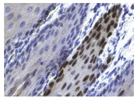
Immunoperoxidase staining of formalin fixed, paraffin-embedded human esophagus tissue showing nuclear staining of squamous epithelial cells.Application
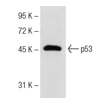
Western blot analysis of p53 expression in Lac-Z whole cell lysate. Antibody tested: p53 (FL-393): .Application
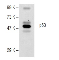
Western blot analysis of p53 expression in Jurkat whole cell lysate. Antibody tested: p53 (FL-393)-G: .Application
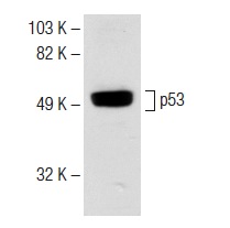
Western blot analysis of p53 expression in Lac-Z whole cell lysate. Antibody tested: p53 (FL-393)-G: .Application
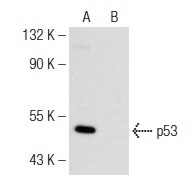
Western blot analysis of p53 expression in untreated (A) and Farnesyl Thiosalicylic Acid Amide treated (B) SW480 whole cell lysates. Note down regulation of p53 expression in lane B.Application
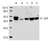
Western blot analysis of p53 expression in A-431 (A), BC3H1 (B), MCP-5 (C), Lac Z (D) and WR19L (E) whole cell lysates.Application
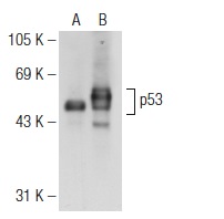
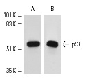
Western blot analysis of p53 expression in non-transfected: (A) and human p53 transfected: (B) 293T whole cell lysates.Application
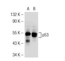
Western blot analysis of p53 expression in non-transfected: (A) and mouse p53 transfected: (B) 293T whole cell lysates.Application
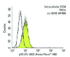
Intracellular FCM analysis of fixed and permeabilized HeLa cells. Black line histogram represents the isotype control, normal goat IgG: .Application
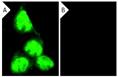
Immunofluorescence staining of methanol-fixed, control HeLa (A) and p53 siRNA silenced HeLa (B) cells showing diminished nuclear staining in the siRNA silenced cells. Cells probed with p53 (FL-393): .Application
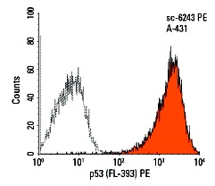
Intracellular FCM analysis of fixed and permeabilized A-431 cells. Black line histogram represents the isotype control, normal rabbit IgG: .Application
Western blot analysis of p53 expression in A-431 (A), WR19L (B) and mouse Lac Z (C) whole cell lysates.Application
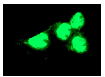
Immunofluorescence staining of methanol-fixed A-431 cells showing nuclear localization.Application
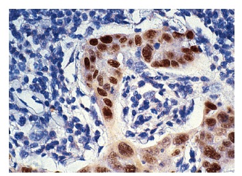
Immunoperoxidase staining of formalin-fixed, paraffin-embedded human breast carcinoma tissue. Note nuclear staining of ductal epithelial.| Product Name | p53 Antibody (FL-393) |
|---|---|
| Antibody Type | Primary Antibodies |
| Modification Notes | p53, a DNA-binding, oligomerization domain- and transcription activation domain-containing tumor suppressor, upregulates growth arrest and apoptosis-related genes in response to stress signals, thereby influencing programmed cell death, cell differentiation, and cell cycle control mechanisms. p53 localizes to the nucleus, yet can be chaperoned to the cytoplasm by the negative regulator, MDM2. MDM2 is an E3 ubiquitin ligase that is upregulated in the presence of active p53, where it poly-ubiquitinates p53 for proteasome targeting. p53 fluctuates between latent and active DNA-binding conformations and is differentially activated through posttranslational modifications, including phosphorylation and acetylation. Mutations in the DNA-binding domain (DBD) of p53, amino acids 110-286, can compromise energetically-favorable association with cis elements and are implicated in several human cancers. |
| Clonality | Polyclonal |
|---|---|
| Isotype | IgG |
| Host Species | Rabbit |
| Tested Applications | WBIPIFIHCFCELISA |
| recommended for detection of p53 of mouse, rat and human origin by WB, IP, IF, IHC(P), FCM and ELISA | |
| Species Reactivity | HumanMouseRat |
| Concentration | 1mg/ml |
| Purification | Affinity purified |
| Alternative Names | epitope mapping within amino acids 1-393 representing full length p53 of human origin |
|---|---|
| Tissue Specificity | epitope mapping within amino acids 1-393 representing full length p53 of human origin |
| Entrez Gene | 7157 |
|---|
Application
Immunoperoxidase staining of formalin fixed, paraffin-embedded human esophagus tissue showing nuclear staining of squamous epithelial cells.
Application
Western blot analysis of p53 expression in Lac-Z whole cell lysate. Antibody tested: p53 (FL-393): .
Application
Western blot analysis of p53 expression in Jurkat whole cell lysate. Antibody tested: p53 (FL-393)-G: .
Application
Western blot analysis of p53 expression in Lac-Z whole cell lysate. Antibody tested: p53 (FL-393)-G: .
Application
Western blot analysis of p53 expression in untreated (A) and Farnesyl Thiosalicylic Acid Amide treated (B) SW480 whole cell lysates. Note down regulation of p53 expression in lane B.
Application
Western blot analysis of p53 expression in A-431 (A), BC3H1 (B), MCP-5 (C), Lac Z (D) and WR19L (E) whole cell lysates.
Application
Western blot analysis of p53 expression in non-transfected: (A) and human p53 transfected: (B) 293T whole cell lysates.
Application
Western blot analysis of p53 expression in non-transfected: (A) and mouse p53 transfected: (B) 293T whole cell lysates.
Application
Intracellular FCM analysis of fixed and permeabilized HeLa cells. Black line histogram represents the isotype control, normal goat IgG: .
Application
Immunofluorescence staining of methanol-fixed, control HeLa (A) and p53 siRNA silenced HeLa (B) cells showing diminished nuclear staining in the siRNA silenced cells. Cells probed with p53 (FL-393): .
Application
Intracellular FCM analysis of fixed and permeabilized A-431 cells. Black line histogram represents the isotype control, normal rabbit IgG: .
Application
Western blot analysis of p53 expression in A-431 (A), WR19L (B) and mouse Lac Z (C) whole cell lysates.
Application
Immunofluorescence staining of methanol-fixed A-431 cells showing nuclear localization.
Application
Immunoperoxidase staining of formalin-fixed, paraffin-embedded human breast carcinoma tissue. Note nuclear staining of ductal epithelial.| Application Notes | recommended for detection of p53 of mouse, rat and human origin by WB, IP, IF, IHC(P), FCM and ELISA |
|---|
| Form | Liquid |
|---|---|
| Storage Instructions | For short-term storage, store at 4° C. For long-term storage, aliquot and store at -20ºC or below. Avoid multiple freeze-thaw cycles. |
| Storage Buffer | phosphate buffered saline , pH 7.4, 150mM NaCl, 0.02% sodium azide and 50% glycerol. |


