Application
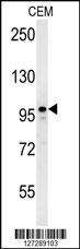
MAP1S Antibody (Center) western blot analysis in CEM cell line lysates (35ug/lane).This demonstrates the MAP1S antibody detected the MAP1S protein (arrow).Application
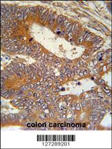
MAP1S Antibody (Center) immunohistochemistry analysis in formalin fixed and paraffin embedded human colon carcinoma followed by peroxidase conjugation of the secondary antibody and DAB staining. This data demonstrates the use of the MAP1S Antibody (Center) for immunohistochemistry. Clinical relevance has not been evaluated.Application
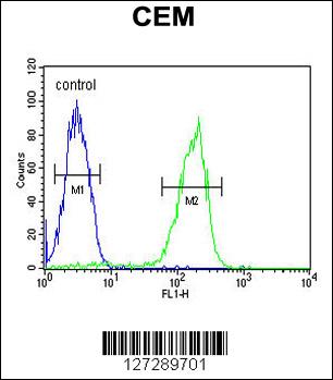
MAP1S Antibody (Center) flow cytometric analysis of CEM cells (right histogram) compared to a negative control cell (left histogram).FITC-conjugated goat-anti-rabbit secondary antibodies were used for the analysis.| Product Name | MAP1S Antibody (Center) |
|---|---|
| Antibody Type | Primary Antibodies |
| Antigen Alias | MAP1S; BPY2IP1; C19orf5; MAP8; VCY2IP1; Microtubule-associated protein 1S; BPY2-interacting protein 1; Microtubule-associated protein 8; Variable charge Y chromosome 2-interacting protein 1; MAP1S heavy chain; MAP1S light chain |
| Clonality | Polyclonal |
|---|---|
| Isotype | Ig |
| Host Species | Rabbit |
| Tested Applications | WBIHCFC |
| WB:1:100~500 IHC |
|
| Species Reactivity | Human |
| Concentration | 1mg/ml |
| Gene Synonyms | BPY2IP1 C19orf5 MAP8 VCY2IP1 |
|---|---|
| Alternative Names | MAP1S BPY2IP1 C19orf5 MAP8 VCY2IP1 Microtubule-associated protein 1S BPY2-interacting protein 1 Microtubule-associated protein 8 Variable charge Y chromosome 2-interacting protein 1 MAP1S heavy chain MAP1S light chain |
| Molecular Weight(MW) | 112211 Da |
| Function | Microtubule-associated protein that mediates aggregation of mitochondria resulting in cell death and genomic destruction (MAGD). Plays a role in anchoring the microtubule-organizing center to the centrosomes. Binds to DNA. Plays a role in apoptosis. Involved in the formation of microtubule bundles (By similarity) |
| Tissue Specificity | Expressed in neurons (at protein level). Expressed in spermatocytes, spermatids and spermatozoa. Expressed in the cerebral cortex. Highly expressed in testis. Moderately expressed in the brain, colon, heart, kidney, liver, lung, placenta, small intestine, spleen and stomach. Weakly expressed in muscle |
| Cellular Localization | Nucleus. Cytoplasm, cytosol. Cytoplasm, cytoskeleton. Cytoplasm, cytoskeleton, spindle. Note=Detected in filopodia-like protrusions and synapses (By similarity). Detected in perinuclear punctate network corresponding to mitochondrial aggregates and in the nucleus in cells exhibiting apoptosis Associated specifically with microtubules stabilized by paclitaxel and colocalizes with RASSF1 isoform A. In interphase cells, shows a diffuse cytoplasmic staining with partial localization to the microtubules. During the different stages of mitosis detected at the spindle microtubules |
| Entrez Gene | 55201 |
|---|---|
| Nucleotide Accession | NP_060644.4 |
Application
MAP1S Antibody (Center) western blot analysis in CEM cell line lysates (35ug/lane).This demonstrates the MAP1S antibody detected the MAP1S protein (arrow).
Application
MAP1S Antibody (Center) immunohistochemistry analysis in formalin fixed and paraffin embedded human colon carcinoma followed by peroxidase conjugation of the secondary antibody and DAB staining. This data demonstrates the use of the MAP1S Antibody (Center) for immunohistochemistry. Clinical relevance has not been evaluated.
Application
MAP1S Antibody (Center) flow cytometric analysis of CEM cells (right histogram) compared to a negative control cell (left histogram).FITC-conjugated goat-anti-rabbit secondary antibodies were used for the analysis.| Application Notes | WB:1:100~500 IHC |
|---|
| Form | Liquid |
|---|---|
| Storage Instructions | For short-term storage, store at 4° C. For long-term storage, aliquot and store at -20ºC or below. Avoid multiple freeze-thaw cycles. |
| Storage Buffer | Purified polyclonal antibody supplied in PBS with 0.09% (W/V) sodium azide. This antibody is purified through a protein A column, followed by peptide affinity purification. |


