Application
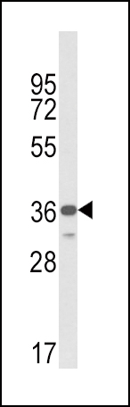
Western blot analysis of MICA Antibody (Center) in MDA-MB231 cell line lysates (35ug/lane). MICA (arrow) was detected using the purified Pab.Application
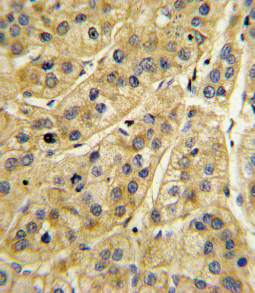
Formalin-fixed and paraffin-embedded human hepatocarcinoma with MICA Antibody (Center), which was peroxidase-conjugated to the secondary antibody, followed by DAB staining. This data demonstrates the use of this antibody for immunohistochemistry; clinical relevance has not been evaluated.Application
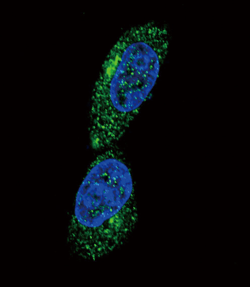
Confocal immunofluorescent analysis of MICA Antibody (Center)(Cat#AP8626c) with MDA-MB231 cell followed by Alexa Fluor搴?488-conjugated goat anti-rabbit lgG (green). DAPI was used to stain the cell nuclear (blue).Application
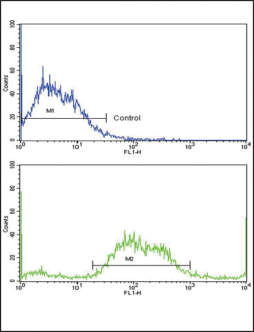
Flow cytometric analysis of SK-Br-3 cells using MICA Antibody (Center)(bottom histogram) compared to a negative control cell (top histogram). FITC-conjugated goat-anti-rabbit secondary antibodies were used for the analysis.| Product Name | MICA Antibody (Center) |
|---|---|
| Antibody Type | Primary Antibodies |
| Antigen Alias | MICA; PERB11.1; MHC class I polypeptide-related sequence A |
| Clonality | Polyclonal |
|---|---|
| Isotype | Ig |
| Host Species | Rabbit |
| Tested Applications | WBIHCIFFC |
| WB:1:100~500 IHC |
|
| Species Reactivity | Human |
| Concentration | 1mg/ml |
| Gene Synonyms | PERB11.1 |
|---|---|
| Alternative Names | MICA PERB11.1 MHC class I polypeptide-related sequence A |
| Molecular Weight(MW) | 42915 Da |
| Function | Seems to have no role in antigen presentation. Acts as a stress-induced self-antigen that is recognized by gamma delta T- cells. Ligand for the KLRK1/NKG2D receptor. Binding to KLRK1 leads to cell lysis |
| Tissue Specificity | Widely expressed with the exception of the central nervous system where it is absent. Expressed predominantly in gastric epithelium and also in monocytes, keratinocytes, endothelial cells, fibroblasts and in the outer layer of Hassal's corpuscles within the medulla of normal thymus. In skin, expressed mainly in the keratin layers, basal cells, ducts and follicles Also expressed in many, but not all, epithelial tumors of lung, breast, kidney, ovary, prostate and colon. In thyomas, overexpressed in cortical and medullar epithelial cells. Tumors expressing MICA display increased levels of gamma delta T-cells |
| Cellular Localization | Cell membrane; Single-pass type I membrane protein. Cytoplasm. Note=Expressed on the cell surface in gastric epithelium, endothelial cells and fibroblasts and in the cytoplasm in keratinocytes and monocytes. Infection with human adenovirus 5 suppresses cell surface expression due to the adenoviral E3-19K protein which causes retention in the endoplasmic reticulum |
| Entrez Gene | 100507436 |
|---|
Application
Western blot analysis of MICA Antibody (Center) in MDA-MB231 cell line lysates (35ug/lane). MICA (arrow) was detected using the purified Pab.
Application
Formalin-fixed and paraffin-embedded human hepatocarcinoma with MICA Antibody (Center), which was peroxidase-conjugated to the secondary antibody, followed by DAB staining. This data demonstrates the use of this antibody for immunohistochemistry; clinical relevance has not been evaluated.
Application
Confocal immunofluorescent analysis of MICA Antibody (Center)(Cat#AP8626c) with MDA-MB231 cell followed by Alexa Fluor搴?488-conjugated goat anti-rabbit lgG (green). DAPI was used to stain the cell nuclear (blue).
Application
Flow cytometric analysis of SK-Br-3 cells using MICA Antibody (Center)(bottom histogram) compared to a negative control cell (top histogram). FITC-conjugated goat-anti-rabbit secondary antibodies were used for the analysis.| Application Notes | WB:1:100~500 IHC |
|---|
| Form | Liquid |
|---|---|
| Storage Instructions | For short-term storage, store at 4° C. For long-term storage, aliquot and store at -20ºC or below. Avoid multiple freeze-thaw cycles. |
| Storage Buffer | Purified polyclonal antibody supplied in PBS with 0.09% (W/V) sodium azide. This antibody is purified through a protein A column, followed by peptide affinity purification. |


