Application
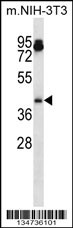
Mouse Aurka Antibody (N-term) western blot analysis in mouse NIH-3T3 cell line lysates (35ug/lane).This demonstrates the Aurka antibody detected the Aurka protein (arrow).| Product Name | Aurka Antibody (N-term) |
|---|---|
| Antibody Type | Primary Antibodies |
| Antigen Alias | Aurka; Aik; Airk; Ark1; Aura; Ayk1; Btak; Iak1; Stk15; Stk6; Aurora kinase A; Aurora 2; Aurora family kinase 1; Aurora/IPL1-related kinase 1; Ipl1- and aurora-related kinase 1; Serine/threonine-protein kinase 6; Serine/threonine-protein kinase Ayk1; Serine |
| Clonality | Polyclonal |
|---|---|
| Isotype | Ig |
| Host Species | Rabbit |
| Tested Applications | WB |
| WB:1:100~500 |
|
| Species Reactivity | Human |
| Concentration | 1mg/ml |
| Alternative Names | Aurka Aik Airk Ark1 Aura Ayk1 Btak Iak1 Stk15 Stk6 Aurora kinase A Aurora 2 Aurora family kinase 1 Aurora/IPL1-related kinase 1 Ipl1- and aurora-related kinase 1 Serine/threonine-protein kinase 6 Serine/threonine-protein kinase Ayk1 Serine |
|---|---|
| Molecular Weight(MW) | 44772 Da |
| Function | Mitotic serine/threonine kinases that contributes to the regulation of cell cycle progression. Associates with the centrosome and the spindle microtubules during mitosis and plays a critical role in various mitotic events including the establishment of mitotic spindle, centrosome duplication, centrosome separation as well as maturation, chromosomal alignment, spindle assembly checkpoint, and cytokinesis. Required for initial activation of CDK1 at centrosomes. Phosphorylates numerous target proteins, including ARHGEF2, BORA, BRCA1, CDC25B, DLGP5, HDAC6, KIF2A, LATS2, NDEL1, PARD3, PPP1R2, PLK1, RASSF1, TACC3, p53/TP53 and TPX2. Regulates KIF2A tubulin depolymerase activity. Required for normal axon formation. Plays a role in microtubule remodeling during neurite extension. Important for microtubule formation and/or stabilization. Also acts as a key regulatory component of the p53/TP53 pathway, and particularly the checkpoint-response pathways critical for oncogenic transformation of cells, by phosphorylating and stabilizing p53/TP53 Phosphorylates its own inhibitors, the protein phosphatase type 1 (PP1) isoforms, to inhibit their activity. Necessary for proper cilia disassembly prior to mitosis |
| Tissue Specificity | Detected in embryonic neurons in dorsal root ganglia and brain cortex (at protein level). Highly expressed in testis, in about one third of the seminiferous tubules. Expression is restricted to specific spermatocytes nearing completion of prophase, with levels falling off on transition to elongated spermatids. Highly expressed in the ovary, expression in the oocyte starts around the transition to large growing follicle Abundant expression is seen in the proliferating granulosa and thecal cells of the growing follicle, and in the young corpus luteum. Very weakly expressed in spleen and intestine |
| Cellular Localization | Cytoplasm, cytoskeleton, centrosome. Cytoplasm, cytoskeleton, spindle pole. Note=Localizes on centrosomes in interphase cells and at each spindle pole in mitosis. Associates with both the pericentriolar material (PCM) and centrioles (By similarity). Detected at the neurite hillock in developing neurons |
| Entrez Gene | 20878 |
|---|---|
| Nucleotide Accession | NP_035627.1 |
Application
Mouse Aurka Antibody (N-term) western blot analysis in mouse NIH-3T3 cell line lysates (35ug/lane).This demonstrates the Aurka antibody detected the Aurka protein (arrow).| Application Notes | WB:1:100~500 |
|---|
| Form | Liquid |
|---|---|
| Storage Instructions | For short-term storage, store at 4° C. For long-term storage, aliquot and store at -20ºC or below. Avoid multiple freeze-thaw cycles. |
| Storage Buffer | Purified polyclonal antibody supplied in PBS with 0.09% (W/V) sodium azide. This antibody is purified through a protein A column, followed by peptide affinity purification. |


