Application
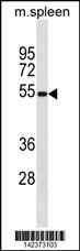
MOUSE Mavs Antibody (N-term) western blot analysis in mouse spleen tissue lysates (35ug/lane).This demonstrates the MOUSE Mavs antibody detected the MOUSE Mavs protein (arrow).| Product Name | Mavs Antibody (N-term) |
|---|---|
| Antibody Type | Primary Antibodies |
| Antigen Alias | Mavs; Ips1; Visa; Mitochondrial antiviral-signaling protein; CARD adapter inducing interferon beta; Interferon beta promoter stimulator protein 1; Virus-induced-signaling adapter |
| Clonality | Polyclonal |
|---|---|
| Isotype | Ig |
| Host Species | Rabbit |
| Tested Applications | WB |
| WB:1:100~500 |
|
| Species Reactivity | Human |
| Concentration | 1mg/ml |
| Gene Synonyms | Ips1 Visa |
|---|---|
| Alternative Names | Mavs Ips1 Visa Mitochondrial antiviral-signaling protein CARD adapter inducing interferon beta Interferon beta promoter stimulator protein 1 Virus-induced-signaling adapter |
| Molecular Weight(MW) | 53399 Da |
| Function | Required for innate immune defense against viruses. Acts downstream of DDX58/RIG-I and IFIH1/MDA5, which detect intracellular dsRNA produced during viral replication, to coordinate pathways leading to the activation of NF-kappa-B, IRF3 and IRF7, and to the subsequent induction of antiviral cytokines such as IFN-beta and RANTES (CCL5). Peroxisomal and mitochondrial MAVS act sequentially to create an antiviral cellular state. Upon viral infection, peroxisomal MAVS induces the rapid interferon- independent expression of defense factors that provide short-term protection, whereas mitochondrial MAVS activates an interferon- dependent signaling pathway with delayed kinetics, which amplifies and stabilizes the antiviral response. May activate the same pathways following detection of extracellular dsRNA by TLR3. May protect cells from apoptosis (By similarity) |
| Tissue Specificity | This MOUSE Mavs antibody is generated from rabbits immunized with a KLH conjugated synthetic peptide between 1-30 amino acids from the N-terminal region of mouse Mavs. |
| Cellular Localization | Mitochondrion outer membrane (By similarity). Mitochondrion (By similarity). Peroxisome (By similarity) |
| Entrez Gene | 228607 |
|---|---|
| Nucleotide Accession | NP_659137.1 |
Application
MOUSE Mavs Antibody (N-term) western blot analysis in mouse spleen tissue lysates (35ug/lane).This demonstrates the MOUSE Mavs antibody detected the MOUSE Mavs protein (arrow).| Application Notes | WB:1:100~500 |
|---|
| Form | Liquid |
|---|---|
| Storage Instructions | For short-term storage, store at 4° C. For long-term storage, aliquot and store at -20ºC or below. Avoid multiple freeze-thaw cycles. |
| Storage Buffer | Purified polyclonal antibody supplied in PBS with 0.09% (W/V) sodium azide. This antibody is purified through a protein A column, followed by peptide affinity purification. |


