Application
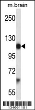
Mouse Pdgfra Antibody (Center) western blot analysis in mouse brain tissue lysates (35ug/lane).This demonstrates the Pdgfra antibody detected the Pdgfra protein (arrow).| Product Name | Pdgfra Antibody (Center) |
|---|---|
| Antibody Type | Primary Antibodies |
| Clonality | Polyclonal |
|---|---|
| Isotype | Ig |
| Host Species | Rabbit |
| Tested Applications | WB |
| WB:1:100~500 |
|
| Species Reactivity | Human |
| Concentration | 1mg/ml |
| Alternative Names | Pdgfra Platelet-derived growth factor receptor alpha Alpha platelet-derived growth factor receptor Alpha-type platelet-derived growth factor receptor CD140 antigen-like family member A Platelet-derived growth factor alpha receptor CD_antigen=CD140a |
|---|---|
| Molecular Weight(MW) | 122683 Da |
| Function | Tyrosine-protein kinase that acts as a cell-surface receptor for PDGFA, PDGFB and PDGFC and plays an essential role in the regulation of embryonic development, cell proliferation, survival and chemotaxis. Depending on the context, promotes or inhibits cell proliferation and cell migration. Plays an important role in the differentiation of bone marrow-derived mesenchymal stem cells. Required for normal skeleton development and cephalic closure during embryonic development. Required for normal development of the mucosa lining the gastrointestinal tract, and for recruitment of mesenchymal cells and normal development of intestinal villi. Plays a role in cell migration and chemotaxis in wound healing. Plays a role in platelet activation, secretion of agonists from platelet granules, and in thrombin-induced platelet aggregation. Binding of its cognate ligands - homodimeric PDGFA, homodimeric PDGFB, heterodimers formed by PDGFA and PDGFB or homodimeric PDGFC -leads to the activation of several signaling cascades; the response depends on the nature of the bound ligand and is modulated by the formation of heterodimers between PDGFRA and PDGFRB. Phosphorylates PIK3R1, PLCG1, and PTPN11. Activation of PLCG1 leads to the production of the cellular signaling molecules diacylglycerol and inositol 1,4,5-trisphosphate, mobilization of cytosolic Ca(2+) and the activation of protein kinase C. Phosphorylates PIK3R1, the regulatory subunit of phosphatidylinositol 3-kinase, and thereby mediates activation of the AKT1 signaling pathway. Mediates activation of HRAS and of the MAP kinases MAPK1/ERK2 and/or MAPK3/ERK1. Promotes activation of STAT family members STAT1, STAT3 and STAT5A and/or STAT5B Receptor signaling is down-regulated by protein phosphatases that dephosphorylate the receptor and its down-stream effectors, and by rapid internalization of the activated receptor |
| Tissue Specificity | Focally expressed in cortical interstitial cells and highly expressed in the interstitium of the papillary region. Also expressed by adventitial cells in arterial vessels Up-regulated in areas of renal fibrosis. In mice with unilateral ureteral obstruction, expression in cortical interstitial cells becomes prominent at day 4 which increases progressively until day 14 |
| Cellular Localization | Cell membrane; Single-pass type I membrane protein. Note=The activated receptor is rapidly internalized and degraded (By similarity) |
| Entrez Gene | 18595 |
|---|---|
| Nucleotide Accession | NP_035188.2 NP_001076785.1 |
Application
Mouse Pdgfra Antibody (Center) western blot analysis in mouse brain tissue lysates (35ug/lane).This demonstrates the Pdgfra antibody detected the Pdgfra protein (arrow).| Application Notes | WB:1:100~500 |
|---|
| Form | Liquid |
|---|---|
| Storage Instructions | For short-term storage, store at 4° C. For long-term storage, aliquot and store at -20ºC or below. Avoid multiple freeze-thaw cycles. |
| Storage Buffer | Purified polyclonal antibody supplied in PBS with 0.09% (W/V) sodium azide. This antibody is purified through a protein A column, followed by peptide affinity purification. |


