Application
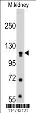
Western blot analysis of anti-MUSK Pab in mouse kidney tissue lysates (35ug/lane). MUSK(arrow) was detected using the purified Pab (1:60 dilution).Application
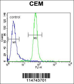
MUSK Antibody flow cytometric analysis of CEM cells (right histogram) compared to a negative control cell (left histogram).FITC-conjugated goat-anti-rabbit secondary antibodies were used for the analysis.Application
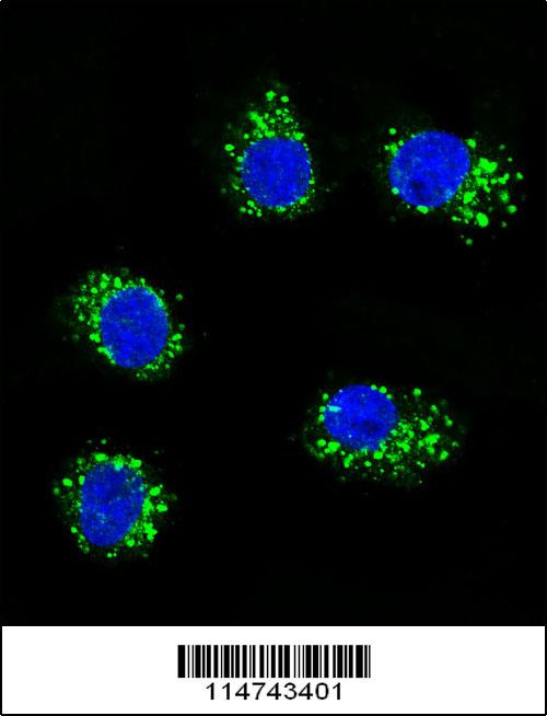
Confocal immunofluorescent analysis of MUSK Antibody(Cat#AP7664d) with MDA-MB231 cell followed by Alexa Fluor 488-conjugated goat anti-rabbit lgG (green).DAPI was used to stain the cell nuclear (blue).| Product Name | MUSK Antibody |
|---|---|
| Antibody Type | Primary Antibodies |
| Antigen Alias | MUSK; Muscle, skeletal receptor tyrosine-protein kinase; Muscle-specific tyrosine-protein kinase receptor |
| Clonality | Polyclonal |
|---|---|
| Isotype | Ig |
| Host Species | Rabbit |
| Tested Applications | WBFCIF |
| WB:1:50~100 FC |
|
| Species Reactivity | Human |
| Concentration | 1mg/ml |
| Alternative Names | MUSK Muscle skeletal receptor tyrosine-protein kinase Muscle-specific tyrosine-protein kinase receptor |
|---|---|
| Molecular Weight(MW) | 97056 Da |
| Function | Receptor tyrosine kinase which plays a central role in the formation and the maintenance of the neuromuscular junction (NMJ), the synapse between the motor neuron and the skeletal muscle. Recruitment of AGRIN by LRP4 to the MUSK signaling complex induces phosphorylation and activation of MUSK, the kinase of the complex. The activation of MUSK in myotubes regulates the formation of NMJs through the regulation of different processes including the specific expression of genes in subsynaptic nuclei, the reorganization of the actin cytoskeleton and the clustering of the acetylcholine receptors (AChR) in the postsynaptic membrane May regulate AChR phosphorylation and clustering through activation of ABL1 and Src family kinases which in turn regulate MUSK. DVL1 and PAK1 that form a ternary complex with MUSK are also important for MUSK-dependent regulation of AChR clustering. May positively regulate Rho family GTPases through FNTA. Mediates the phosphorylation of FNTA which promotes prenylation, recruitment to membranes and activation of RAC1 a regulator of the actin cytoskeleton and of gene expression. Other effectors of the MUSK signaling include DNAJA3 which functions downstream of MUSK. May also play a role within the central nervous system by mediating cholinergic responses, synaptic plasticity and memory formation (By similarity) |
| Tissue Specificity | This MUSK antibody is generated from rabbits immunized with human recombinant MUSK protein. |
| Cellular Localization | Cell junction, synapse, postsynaptic cell membrane; Single-pass type I membrane protein (Probable) Note=Localizes to the postsynaptic cell membrane of the neuromuscular junction (Probable) |
| Entrez Gene | 4593 |
|---|
Application
Western blot analysis of anti-MUSK Pab in mouse kidney tissue lysates (35ug/lane). MUSK(arrow) was detected using the purified Pab (1:60 dilution).
Application
MUSK Antibody flow cytometric analysis of CEM cells (right histogram) compared to a negative control cell (left histogram).FITC-conjugated goat-anti-rabbit secondary antibodies were used for the analysis.
Application
Confocal immunofluorescent analysis of MUSK Antibody(Cat#AP7664d) with MDA-MB231 cell followed by Alexa Fluor 488-conjugated goat anti-rabbit lgG (green).DAPI was used to stain the cell nuclear (blue).| Application Notes | WB:1:50~100 FC |
|---|
| Form | Liquid |
|---|---|
| Storage Instructions | For short-term storage, store at 4° C. For long-term storage, aliquot and store at -20ºC or below. Avoid multiple freeze-thaw cycles. |
| Storage Buffer | Purified polyclonal antibody supplied in PBS with 0.09% (W/V) sodium azide. This antibody is prepared by Saturated Ammonium Sulfate (SAS) precipitation followed by dialysis against PBS. |


