Application
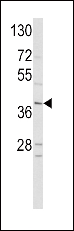
Western blot analysis of hNeuroD1-Q30 in HepG2 cell line lysates (35ug/lane). NEUROD1 (arrow) was detected using the purified Pab.Application
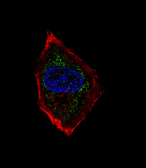
Fluorescent confocal image of HepG2 cell stained with hNeuroD1-Q30(Cat#AP2021a). HepG2 cells were fixed with 4% PFA (20 min), permeabilized with Triton X-100 (0.1%, 10 min), then incubated with hNeuroD1-Q30 primary antibody (1:25, 1 h at 37℃). For secondary antibody, Alexa Fluor® 488 conjugated donkey anti-rabbit antibody (green) was used (1:400, 50 min at 37℃).Cytoplasmic actin was counterstained with Alexa Fluor® 555 (red) conjugated Phalloidin (7units/ml, 1 h at 37℃). Nuclei were counterstained with DAPI (blue) (10 µg/ml, 10 min).hNeuroD1-Q30 immunoreactivity is localized to vesicles significantly.| Product Name | NeuroD1 Antibody (N-term) |
|---|---|
| Antibody Type | Primary Antibodies |
| Antigen Alias | NEUROD1; BHLHA3; NEUROD; Neurogenic differentiation factor 1; Class A basic helix-loop-helix protein 3 |
| Clonality | Polyclonal |
|---|---|
| Isotype | Ig |
| Host Species | Rabbit |
| Tested Applications | WBIF |
| WB:1:100~500 IF |
|
| Species Reactivity | Human |
| Concentration | 1mg/ml |
| Gene Synonyms | BHLHA3 NEUROD |
|---|---|
| Alternative Names | NEUROD1 BHLHA3 NEUROD Neurogenic differentiation factor 1 Class A basic helix-loop-helix protein 3 |
| Molecular Weight(MW) | 39920 Da |
| Function | Acts as a transcriptional activator: mediates transcriptional activation by binding to E box-containing promoter consensus core sequences 5'-CANNTG-3'. Associates with the p300/CBP transcription coactivator complex to stimulate transcription of the secretin gene as well as the gene encoding the cyclin-dependent kinase inhibitor CDKN1A. Contributes to the regulation of several cell differentiation pathways, like those that promote the formation of early retinal ganglion cells, inner ear sensory neurons, granule cells forming either the cerebellum or the dentate gyrus cell layer of the hippocampus, endocrine islet cells of the pancreas and enteroendocrine cells of the small intestine. Together with PAX6 or SIX3, is required for the regulation of amacrine cell fate specification. Also required for dendrite morphogenesis and maintenance in the cerebellar cortex Associates with chromatin to enhancer regulatory elements in genes encoding key transcriptional regulators of neurogenesis (By similarity) |
| Tissue Specificity | This NeuroD1 antibody is generated from rabbits immunized with a KLH conjugated synthetic peptide between 23~52 amino acids from the N-terminal region of human NeuroD1. |
| Cellular Localization | Cytoplasm (By similarity). Nucleus. Note=In pancreatic islet cells, shuttles to the nucleus in response to glucose stimulation (By similarity). Colocalizes with NR0B2 in the nucleus |
| Entrez Gene | 4760 |
|---|
Application
Western blot analysis of hNeuroD1-Q30 in HepG2 cell line lysates (35ug/lane). NEUROD1 (arrow) was detected using the purified Pab.
Application
Fluorescent confocal image of HepG2 cell stained with hNeuroD1-Q30(Cat#AP2021a). HepG2 cells were fixed with 4% PFA (20 min), permeabilized with Triton X-100 (0.1%, 10 min), then incubated with hNeuroD1-Q30 primary antibody (1:25, 1 h at 37℃). For secondary antibody, Alexa Fluor® 488 conjugated donkey anti-rabbit antibody (green) was used (1:400, 50 min at 37℃).Cytoplasmic actin was counterstained with Alexa Fluor® 555 (red) conjugated Phalloidin (7units/ml, 1 h at 37℃). Nuclei were counterstained with DAPI (blue) (10 µg/ml, 10 min).hNeuroD1-Q30 immunoreactivity is localized to vesicles significantly.| Application Notes | WB:1:100~500 IF |
|---|
| Form | Liquid |
|---|---|
| Storage Instructions | For short-term storage, store at 4° C. For long-term storage, aliquot and store at -20ºC or below. Avoid multiple freeze-thaw cycles. |
| Storage Buffer | Purified polyclonal antibody supplied in PBS with 0.09% (W/V) sodium azide. This antibody is purified through a protein G column, eluted with high and low pH buffers and neutralized immediately, followed by dialysis against PBS. |


