Application
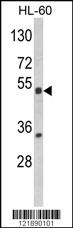
Western blot analysis of NR1I2 Antibody (Center) in HL-60 cell line lysates (35ug/lane). NR1I2 (arrow) was detected using the purified Pab.Application
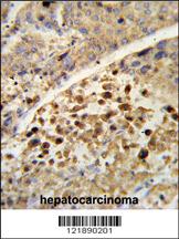
Formalin-fixed and paraffin-embedded human hepatocarcinoma reacted with NR1I2 Antibody (Center), which was peroxidase-conjugated to the secondary antibody, followed by DAB staining. This data demonstrates the use of this antibody for immunohistochemistry; clinical relevance has not been evaluated.Application
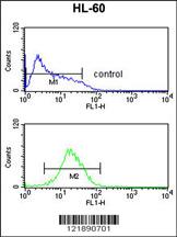
NR1I2 Antibody (Center) flow cytometric analysis of HL-60 cells (bottom histogram) compared to a negative control cell (top histogram).FITC-conjugated goat-anti-rabbit secondary antibodies were used for the analysis.Application
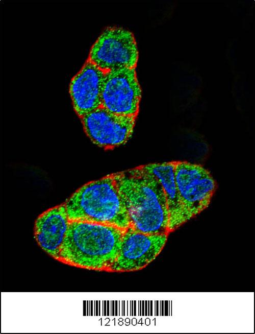
Confocal immunofluorescent analysis of NR1I2 Antibody (Center)(Cat#AP8674c) with HepG2 cell followed by Alexa Fluor 488-conjugated goat anti-rabbit lgG (green). Actin filaments have been labeled with Alexa Fluor 555 phalloidin (red). DAPI was used to stain the cell nuclear (blue).| Product Name | NR1I2 Antibody (Center) |
|---|---|
| Antibody Type | Primary Antibodies |
| Antigen Alias | NR1I2; PXR; Nuclear receptor subfamily 1 group I member 2; Orphan nuclear receptor PAR1; Orphan nuclear receptor PXR; Pregnane X receptor; Steroid and xenobiotic receptor |
| Clonality | Polyclonal |
|---|---|
| Isotype | Ig |
| Host Species | Rabbit |
| Tested Applications | WBIHCFCIF |
| WB:1:100~500 IHC |
|
| Species Reactivity | Human |
| Concentration | 1mg/ml |
| Gene Synonyms | PXR |
|---|---|
| Alternative Names | NR1I2 PXR Nuclear receptor subfamily 1 group I member 2 Orphan nuclear receptor PAR1 Orphan nuclear receptor PXR Pregnane X receptor Steroid and xenobiotic receptor |
| Molecular Weight(MW) | 49762 Da |
| Function | Nuclear receptor that binds and is activated by variety of endogenous and xenobiotic compounds. Transcription factor that activates the transcription of multiple genes involved in the metabolism and secretion of potentially harmful xenobiotics, drugs and endogenous compounds. Activated by the antibiotic rifampicin and various plant metabolites, such as hyperforin, guggulipid, colupulone, and isoflavones. Response to specific ligands is species-specific. Activated by naturally occurring steroids, such as pregnenolone and progesterone. Binds to a response element in the promoters of the CYP3A4 and ABCB1/MDR1 genes |
| Tissue Specificity | Expressed in liver, colon and small intestine. |
| Cellular Localization | Nucleus. |
| Entrez Gene | 8856 |
|---|
Application
Western blot analysis of NR1I2 Antibody (Center) in HL-60 cell line lysates (35ug/lane). NR1I2 (arrow) was detected using the purified Pab.
Application
Formalin-fixed and paraffin-embedded human hepatocarcinoma reacted with NR1I2 Antibody (Center), which was peroxidase-conjugated to the secondary antibody, followed by DAB staining. This data demonstrates the use of this antibody for immunohistochemistry; clinical relevance has not been evaluated.
Application
NR1I2 Antibody (Center) flow cytometric analysis of HL-60 cells (bottom histogram) compared to a negative control cell (top histogram).FITC-conjugated goat-anti-rabbit secondary antibodies were used for the analysis.
Application
Confocal immunofluorescent analysis of NR1I2 Antibody (Center)(Cat#AP8674c) with HepG2 cell followed by Alexa Fluor 488-conjugated goat anti-rabbit lgG (green). Actin filaments have been labeled with Alexa Fluor 555 phalloidin (red). DAPI was used to stain the cell nuclear (blue).| Application Notes | WB:1:100~500 IHC |
|---|
| Form | Liquid |
|---|---|
| Storage Instructions | For short-term storage, store at 4° C. For long-term storage, aliquot and store at -20ºC or below. Avoid multiple freeze-thaw cycles. |
| Storage Buffer | Purified polyclonal antibody supplied in PBS with 0.09% (W/V) sodium azide. This antibody is purified through a protein A column, followed by peptide affinity purification. |


