Application
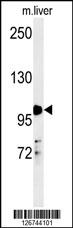
NR3C1 Antibody (Center) western blot analysis in mouse liver tissue lysates (35ug/lane).This demonstrates the NR3C1 antibody detected NR3C1 protein (arrow).Application
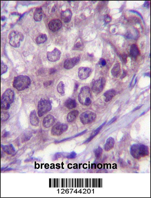
NR3C1 Antibody (Center) immunohistochemistry analysis in formalin fixed and paraffin embedded human breast carcinoma followed by peroxidase conjugation of the secondary antibody and DAB staining.This data demonstrates the use of NR3C1 Antibody (Center) for immunohistochemistry. Clinical relevance has not been evaluated.Application
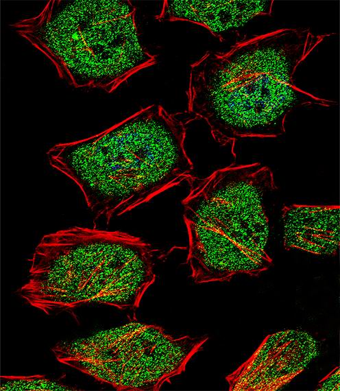
Fluorescent confocal image of Hela cell stained with NR3C1 Antibody (Center)(Cat#AP5468c).Hela cells were fixed with 4% PFA (20 min), permeabilized with Triton X-100 (0.1%, 10 min), then incubated with NR3C1 primary antibody (1:25, 1 h at 37℃). For secondary antibody, Alexa Fluor® 488 conjugated donkey anti-rabbit antibody (green) was used (1:400, 50 min at 37℃).Cytoplasmic actin was counterstained with Alexa Fluor® 555 (red) conjugated Phalloidin (7units/ml, 1 h at 37℃). Nuclei were counterstained with DAPI (blue) (10 µg/ml, 10 min). NR3C1 immunoreactivity is localized to Cytoplasm and Nucleus significantly.| Product Name | NR3C1 Antibody (Center) |
|---|---|
| Antibody Type | Primary Antibodies |
| Antigen Alias | NR3C1; GRL; Glucocorticoid receptor; Nuclear receptor subfamily 3 group C member 1 |
| Clonality | Polyclonal |
|---|---|
| Isotype | Ig |
| Host Species | Rabbit |
| Tested Applications | WBIHCIF |
| WB:1:100~500 IHC |
|
| Species Reactivity | Human |
| Concentration | 1mg/ml |
| Gene Synonyms | GRL |
|---|---|
| Alternative Names | NR3C1 GRL Glucocorticoid receptor Nuclear receptor subfamily 3 group C member 1 |
| Molecular Weight(MW) | 85659 Da |
| Function | Receptor for glucocorticoids (GC). Has a dual mode of action: as a transcription factor that binds to glucocorticoid response elements (GRE), both for nuclear and mitochondrial DNA, and as a modulator of other transcription factors. Affects inflammatory responses, cellular proliferation and differentiation in target tissues. Could act as a coactivator for STAT5-dependent transcription upon growth hormone (GH) stimulation and could reveal an essential role of hepatic GR in the control of body growth. Involved in chromatin remodeling. Plays a significant role in transactivation |
| Tissue Specificity | Widely expressed. In the heart, detected in left and right atria, left and right ventricles, aorta, apex, intraventricular septum, and atrioventricular node as well as whole adult and fetal heart |
| Cellular Localization | Cytoplasm. Mitochondrion. Nucleus. Note=Cytoplasmic in the absence of ligand, nuclear after ligand- binding |
| Entrez Gene | 2908 |
|---|---|
| Nucleotide Accession | NP_000167.1 |
Application
NR3C1 Antibody (Center) western blot analysis in mouse liver tissue lysates (35ug/lane).This demonstrates the NR3C1 antibody detected NR3C1 protein (arrow).
Application
NR3C1 Antibody (Center) immunohistochemistry analysis in formalin fixed and paraffin embedded human breast carcinoma followed by peroxidase conjugation of the secondary antibody and DAB staining.This data demonstrates the use of NR3C1 Antibody (Center) for immunohistochemistry. Clinical relevance has not been evaluated.
Application
Fluorescent confocal image of Hela cell stained with NR3C1 Antibody (Center)(Cat#AP5468c).Hela cells were fixed with 4% PFA (20 min), permeabilized with Triton X-100 (0.1%, 10 min), then incubated with NR3C1 primary antibody (1:25, 1 h at 37℃). For secondary antibody, Alexa Fluor® 488 conjugated donkey anti-rabbit antibody (green) was used (1:400, 50 min at 37℃).Cytoplasmic actin was counterstained with Alexa Fluor® 555 (red) conjugated Phalloidin (7units/ml, 1 h at 37℃). Nuclei were counterstained with DAPI (blue) (10 µg/ml, 10 min). NR3C1 immunoreactivity is localized to Cytoplasm and Nucleus significantly.| Application Notes | WB:1:100~500 IHC |
|---|
| Form | Liquid |
|---|---|
| Storage Instructions | For short-term storage, store at 4° C. For long-term storage, aliquot and store at -20ºC or below. Avoid multiple freeze-thaw cycles. |
| Storage Buffer | Purified polyclonal antibody supplied in PBS with 0.09% (W/V) sodium azide. This antibody is purified through a protein A column, followed by peptide affinity purification. |


