Application
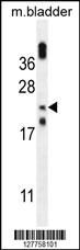
OBFC2A Antibody (Center) western blot analysis in mouse bladder tissue lysates (35ug/lane).This demonstrates the OBFC2A antibody detected the OBFC2A protein (arrow).Application
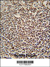
OBFC2A antibody (Center) immunohistochemistry analysis in formalin fixed and paraffin embedded human spleen tissue followed by peroxidase conjugation of the secondary antibody and DAB staining. This data demonstrates the use of the OBFC2A antibody (Center) for immunohistochemistry. Clinical relevance has not been evaluated.Application
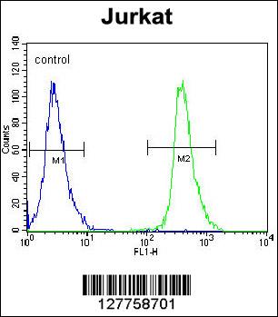
OBFC2A Antibody (Center) flow cytometric analysis of Jurkat cells (right histogram) compared to a negative control cell (left histogram).FITC-conjugated goat-anti-rabbit secondary antibodies were used for the analysis.| Product Name | OBFC2A Antibody (Center) |
|---|---|
| Antibody Type | Primary Antibodies |
| Clonality | Polyclonal |
|---|---|
| Isotype | Ig |
| Host Species | Rabbit |
| Tested Applications | WBIHCFC |
| WB:1:100~500 IHC |
|
| Species Reactivity | Human |
| Concentration | 1mg/ml |
| Gene Synonyms | OBFC2A, SSB2 |
|---|---|
| Alternative Names | NABP1 OBFC2A SSB2 SOSS complex subunit B2 Nucleic acid-binding protein 1 Oligonucleotide/oligosaccharide-binding fold-containing protein 2A Sensor of single-strand DNA complex subunit B2 Sensor of ssDNA subunit B2 Single-stranded DNA-binding protei |
| Molecular Weight(MW) | 22423 Da |
| Function | Component of the SOSS complex, a multiprotein complex that functions downstream of the MRN complex to promote DNA repair and G2/M checkpoint. In the SOSS complex, acts as a sensor of single-stranded DNA that binds to single-stranded DNA, in particular to polypyrimidines. The SOSS complex associates with DNA lesions and influences diverse endpoints in the cellular DNA damage response including cell-cycle checkpoint activation, recombinational repair and maintenance of genomic stability Required for efficient homologous recombination-dependent repair of double-strand breaks (DSBs) and ATM-dependent signaling pathways |
| Tissue Specificity | This OBFC2A antibody is generated from rabbits immunized with a KLH conjugated synthetic peptide between 105-134 amino acids from the Central region of human OBFC2A. |
| Cellular Localization | Nucleus. Note=Localizes to nuclear foci following DNA damage |
| Entrez Gene | 64859 |
|---|---|
| Nucleotide Accession | NP_001026886.1 |
Application
OBFC2A Antibody (Center) western blot analysis in mouse bladder tissue lysates (35ug/lane).This demonstrates the OBFC2A antibody detected the OBFC2A protein (arrow).
Application
OBFC2A antibody (Center) immunohistochemistry analysis in formalin fixed and paraffin embedded human spleen tissue followed by peroxidase conjugation of the secondary antibody and DAB staining. This data demonstrates the use of the OBFC2A antibody (Center) for immunohistochemistry. Clinical relevance has not been evaluated.
Application
OBFC2A Antibody (Center) flow cytometric analysis of Jurkat cells (right histogram) compared to a negative control cell (left histogram).FITC-conjugated goat-anti-rabbit secondary antibodies were used for the analysis.| Application Notes | WB:1:100~500 IHC |
|---|
| Form | Liquid |
|---|---|
| Storage Instructions | For short-term storage, store at 4° C. For long-term storage, aliquot and store at -20ºC or below. Avoid multiple freeze-thaw cycles. |
| Storage Buffer | Purified polyclonal antibody supplied in PBS with 0.09% (W/V) sodium azide. This antibody is purified through a protein A column, followed by peptide affinity purification. |


