Application
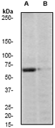
A. Western blot analysis on HeLa cell lysates using anti-Phospho-PAK1 (pS20) RabMAb (Cat. #AJ1583c), 1:2000 dilution. Cells were either (A) untreated (B) treated with AP| Product Name | PAK1 Antibody Phospho (pS20) |
|---|---|
| Antibody Type | Primary Antibodies |
| Antigen Alias | PAK1, Serine/threonine-protein kinase PAK 1, p65-PAK;Alpha-PAK |
| Modification | p-S20 |
| Clonality | Monoclonal |
|---|---|
| Clone Number | EP655Y |
| Host Species | Rabbit |
| Tested Applications | WB |
| WB:1:2000 |
|
| Species Reactivity | Human |
| Concentration | 1mg/ml |
| Alternative Names | PAK1 Serine/threonine-protein kinase PAK 1 p65-PAK Alpha-PAK |
|---|---|
| Molecular Weight(MW) | 60647 Da |
| Function | Protein kinase involved in intracellular signaling pathways downstream of integrins and receptor-type kinases that plays an important role in cytoskeleton dynamics, in cell adhesion, migration, proliferation, apoptosis, mitosis, and in vesicle-mediated transport processes. Can directly phosphorylate BAD and protects cells against apoptosis. Activated by interaction with CDC42 and RAC1. Functions as GTPase effector that links the Rho-related GTPases CDC42 and RAC1 to the JNK MAP kinase pathway Phosphorylates and activates MAP2K1, and thereby mediates activation of downstream MAP kinases. Involved in the reorganization of the actin cytoskeleton, actin stress fibers and of focal adhesion complexes. Phosphorylates the tubulin chaperone TBCB and thereby plays a role in the regulation of microtubule biogenesis and organization of the tubulin cytoskeleton. Plays a role in the regulation of insulin secretion in response to elevated glucose levels. Part of a ternary complex that contains PAK1, DVL1 and MUSK that is important for MUSK-dependent regulation of AChR clustering during the formation of the neuromuscular junction (NMJ). Activity is inhibited in cells undergoing apoptosis, potentially due to binding of CDC2L1 and CDC2L2. Phosphorylates MYL9/MLC2. Phosphorylates RAF1 at 'Ser-338' and 'Ser-339' resulting in: activation of RAF1, stimulation of RAF1 translocation to mitochondria, phosphorylation of BAD by RAF1, and RAF1 binding to BCL2. Phosphorylates SNAI1 at 'Ser-246' promoting its transcriptional repressor activity by increasing its accumulation in the nucleus |
| Tissue Specificity | A phosphor-specific peptide corresponding to residues surrounding Serine 20 on human PAK1 was used as an immunogen. This antibody recognized PAK1 phosphorylated at Ser20. |
| Cellular Localization | Cytoplasm. Cell junction, focal adhesion. Cell membrane. Note=Recruited to the cell membrane by interaction with CDC42 and RAC1. Recruited to focal adhesions upon activation |
| Entrez Gene | 5058 |
|---|
Application
A. Western blot analysis on HeLa cell lysates using anti-Phospho-PAK1 (pS20) RabMAb (Cat. #AJ1583c), 1:2000 dilution. Cells were either (A) untreated (B) treated with AP| Application Notes | WB:1:2000 |
|---|
| Form | Liquid |
|---|---|
| Storage Instructions | For short-term storage, store at 4° C. For long-term storage, aliquot and store at -20ºC or below. Avoid multiple freeze-thaw cycles. |
| Storage Buffer | 50 mM Tris-Glycine (pH 7.4), 0.15 M NaCl, 40% Glycerol, 0.01% sodium azide and 0.05% BSA. |


