Application
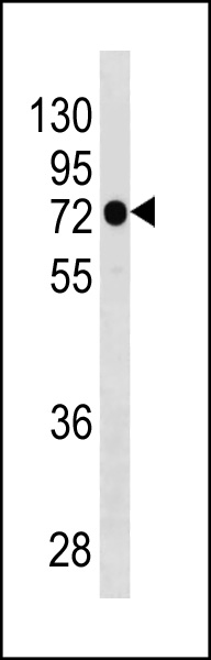
PARP3 Antibody (N-term) western blot analysis in NCI-H292 cell line lysates (35ug/lane).This demonstrates the PARP3 antibody detected the PARP3 protein (arrow).| Product Name | PARP3 Antibody (N-term) |
|---|---|
| Antibody Type | Primary Antibodies |
| Antigen Alias | PARP3;ADPRT3; ADPRTL3; Poly [ADP-ribose] polymerase 3; Poly [ADP-ribose] polymerase 3; ADP-ribosyltransferase diphtheria toxin-like 3; Poly [ADP-ribose] polymerase 3; IRT1; Poly [ADP-ribose] polymerase 3; NAD(+) ADP-ribosyltransferase 3; Poly [ADP-ribose] |
| Clonality | Polyclonal |
|---|---|
| Isotype | Ig |
| Host Species | Rabbit |
| Tested Applications | WB |
| WB:1:100~500 |
|
| Species Reactivity | Human |
| Concentration | 1mg/ml |
| Gene Synonyms | ADPRT3 ADPRTL3 |
|---|---|
| Alternative Names | PARP3 ADPRT3 ADPRTL3 Poly [ADP-ribose] polymerase 3 Poly [ADP-ribose] polymerase 3 ADP-ribosyltransferase diphtheria toxin-like 3 Poly [ADP-ribose] polymerase 3 IRT1 Poly [ADP-ribose] polymerase 3 NAD(+) ADP-ribosyltransferase 3 Poly [ADP-ribose] |
| Molecular Weight(MW) | 60070 Da |
| Function | Involved in the base excision repair (BER) pathway, by catalyzing the poly(ADP-ribosyl)ation of a limited number of acceptor proteins involved in chromatin architecture and in DNA metabolism. This modification follows DNA damages and appears as an obligatory step in a detection/signaling pathway leading to the reparation of DNA strand breaks. May link the DNA damage surveillance network to the mitotic fidelity checkpoint Negatively influences the G1/S cell cycle progression without interfering with centrosome duplication. Binds DNA. May be involved in the regulation of PRC2 and PRC3 complex-dependent gene silencing |
| Tissue Specificity | Widely expressed; the highest levels are in the kidney, skeletal muscle, liver, heart and spleen; also detected in pancreas, lung, placenta, brain, leukocytes, colon, small intestine, ovary, testis, prostate and thymus |
| Cellular Localization | Nucleus. Cytoplasm, cytoskeleton, centrosome. Cytoplasm, cytoskeleton, centrosome, centriole Note=Core component of the centrosome. Preferentially localized to the daughter centriole throughout the cell cycle. According to PubMed:16924674, it is almost exclusively localized in the nucleus and appears in numerous small foci and a small number of larger foci whereas a centrosomal location has not been detected |
| Entrez Gene | 10039 |
|---|
Application
PARP3 Antibody (N-term) western blot analysis in NCI-H292 cell line lysates (35ug/lane).This demonstrates the PARP3 antibody detected the PARP3 protein (arrow).| Application Notes | WB:1:100~500 |
|---|
| Form | Liquid |
|---|---|
| Storage Instructions | For short-term storage, store at 4° C. For long-term storage, aliquot and store at -20ºC or below. Avoid multiple freeze-thaw cycles. |
| Storage Buffer | Purified polyclonal antibody supplied in PBS with 0.09% (W/V) sodium azide. This antibody is purified through a protein A column, followed by peptide affinity purification. |


