Application
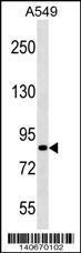
PASD1 Antibody (Center) western blot analysis in A549 cell line lysates (35ug/lane).This demonstrates the PASD1 antibody detected the PASD1 protein (arrow).| Product Name | PASD1 Antibody (Center) |
|---|---|
| Antibody Type | Primary Antibodies |
| Antigen Alias | PASD1; PAS domain-containing protein 1; Cancer/testis antigen 63; OX-TES-1 |
| Clonality | Polyclonal |
|---|---|
| Isotype | Ig |
| Host Species | Rabbit |
| Tested Applications | WB |
| WB:1:100~500 |
|
| Species Reactivity | Human |
| Concentration | 1mg/ml |
| Alternative Names | PASD1 PAS domain-containing protein 1 Cancer/testis antigen 63 OX-TES-1 |
|---|---|
| Molecular Weight(MW) | 87428 Da |
| Function | May function as a transcription factor. |
| Tissue Specificity | Testis-specific. Found in histologically normal tissues from patients with uterus, lung and small intestine cancers. Widespread expression seen in solid tumors and diffuse large B-cell lymphoma (DLBCL)-derived cell lines. Isoform 2 is expressed in all DLBCL-derived cell lines, while isoform 1 is preferentially expressed in cell lines derived from non-germinal center DLBCL |
| Cellular Localization | Nucleus (Probable). |
| Entrez Gene | 139135 |
|---|---|
| Nucleotide Accession | NP_775764.2 |
Application
PASD1 Antibody (Center) western blot analysis in A549 cell line lysates (35ug/lane).This demonstrates the PASD1 antibody detected the PASD1 protein (arrow).| Application Notes | WB:1:100~500 |
|---|
| Form | Liquid |
|---|---|
| Storage Instructions | For short-term storage, store at 4° C. For long-term storage, aliquot and store at -20ºC or below. Avoid multiple freeze-thaw cycles. |
| Storage Buffer | Purified polyclonal antibody supplied in PBS with 0.09% (W/V) sodium azide. This antibody is purified through a protein A column, followed by peptide affinity purification. |


