Application
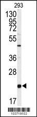
Western blot analysis of anti-PBP Antibody (Center) Antibody (Center) in 293 cell line lysates (35ug/lane). PBP(arrow) was detected using the purified Pab.Application
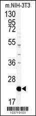
Western blot analysis of anti-PBP Antibody (Center) in mouse NIH-3T3 tissue lysates (35ug/lane). PBP(arrow) was detected using the purified Pab.Application
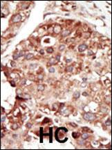
Formalin-fixed and paraffin-embedded human cancer tissue reacted with the primary antibody, which was peroxidase-conjugated to the secondary antibody, followed by DAB staining. This data demonstrates the use of this antibody for immunohistochemistry; clinical relevance has not been evaluated. BC = breast carcinoma; HC = hepatocarcinoma.Application
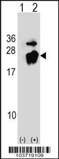
Western blot analysis of PBP (arrow) using rabbit polyclonal PBP Antibody (A152) . 293 cell lysates (2 ug/lane) either nontransfected (Lane 1) or transiently transfected (Lane 2) with the PBP gene.| Product Name | PBP Antibody (Center) |
|---|---|
| Antibody Type | Primary Antibodies |
| Antigen Alias | PEBP1; PBP; PEBP; Phosphatidylethanolamine-binding protein 1; HCNPpp; Neuropolypeptide h3; Prostatic-binding protein; Raf kinase inhibitor protein; Hippocampal cholinergic neurostimulating peptide |
| Clonality | Polyclonal |
|---|---|
| Isotype | Ig |
| Host Species | Rabbit |
| Tested Applications | WBIHC |
| WB:1:100~500 IHC |
|
| Species Reactivity | Human |
| Concentration | 1mg/ml |
| Gene Synonyms | PBP PEBP |
|---|---|
| Alternative Names | PEBP1 PBP PEBP Phosphatidylethanolamine-binding protein 1 HCNPpp Neuropolypeptide h3 Prostatic-binding protein Raf kinase inhibitor protein Hippocampal cholinergic neurostimulating peptide |
| Molecular Weight(MW) | 21057 Da |
| Function | Binds ATP, opioids and phosphatidylethanolamine. Has lower affinity for phosphatidylinositol and phosphatidylcholine Serine protease inhibitor which inhibits thrombin, neuropsin and chymotrypsin but not trypsin, tissue type plasminogen activator and elastase (By similarity). Inhibits the kinase activity of RAF1 by inhibiting its activation and by dissociating the RAF1/MEK complex and acting as a competitive inhibitor of MEK phosphorylation |
| Tissue Specificity | This PBP antibody is generated from rabbits immunized with a KLH conjugated synthetic peptide between 145~175 amino acids from the central region of human PBP. |
| Cellular Localization | Cytoplasm (By similarity). |
| Entrez Gene | 5037 |
|---|
Application
Western blot analysis of anti-PBP Antibody (Center) Antibody (Center) in 293 cell line lysates (35ug/lane). PBP(arrow) was detected using the purified Pab.
Application
Western blot analysis of anti-PBP Antibody (Center) in mouse NIH-3T3 tissue lysates (35ug/lane). PBP(arrow) was detected using the purified Pab.
Application
Formalin-fixed and paraffin-embedded human cancer tissue reacted with the primary antibody, which was peroxidase-conjugated to the secondary antibody, followed by DAB staining. This data demonstrates the use of this antibody for immunohistochemistry; clinical relevance has not been evaluated. BC = breast carcinoma; HC = hepatocarcinoma.
Application
Western blot analysis of PBP (arrow) using rabbit polyclonal PBP Antibody (A152) . 293 cell lysates (2 ug/lane) either nontransfected (Lane 1) or transiently transfected (Lane 2) with the PBP gene.| Application Notes | WB:1:100~500 IHC |
|---|
| Form | Liquid |
|---|---|
| Storage Instructions | For short-term storage, store at 4° C. For long-term storage, aliquot and store at -20ºC or below. Avoid multiple freeze-thaw cycles. |
| Storage Buffer | Purified polyclonal antibody supplied in PBS with 0.09% (W/V) sodium azide. This antibody is purified through a protein G column, eluted with high and low pH buffers and neutralized immediately, followed by dialysis against PBS. |


