Application
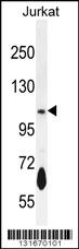
PDE3B Antibody (Center) western blot analysis in Jurkat cell line lysates (35ug/lane).This demonstrates the PDE3B antibody detected the PDE3B protein (arrow).Application
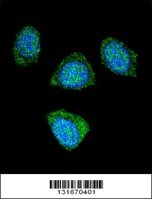
Confocal immunofluorescent analysis of PDE3B Antibody (Center) (Cat#AP12018c) with 293 cell followed by Alexa Fluor 488-conjugated goat anti-rabbit lgG (green). DAPI was used to stain the cell nuclear (blue).Application
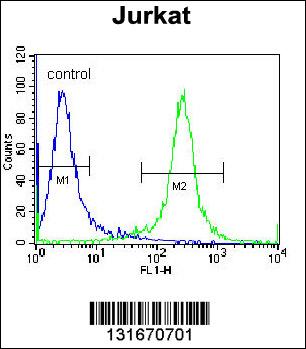
PDE3B Antibody (Center) flow cytometric analysis of Jurkat cells (right histogram) compared to a negative control cell (left histogram).FITC-conjugated donkey-anti-rabbit secondary antibodies were used for the analysis.| Product Name | PDE3B Antibody (Center) |
|---|---|
| Antibody Type | Primary Antibodies |
| Antigen Alias | PDE3B; cGMP-inhibited 3', 5'-cyclic phosphodiesterase B; CGIPDE1; Cyclic GMP-inhibited phosphodiesterase B |
| Clonality | Polyclonal |
|---|---|
| Isotype | Ig |
| Host Species | Rabbit |
| Tested Applications | WBIFFC |
| WB:1:100~500 IF |
|
| Species Reactivity | Human |
| Concentration | 1mg/ml |
| Alternative Names | PDE3B cGMP-inhibited 3' 5'-cyclic phosphodiesterase B CGIPDE1 Cyclic GMP-inhibited phosphodiesterase B |
|---|---|
| Molecular Weight(MW) | 124333 Da |
| Function | Cyclic nucleotide phosphodiesterase with a dual- specificity for the second messengers cAMP and cGMP, which are key regulators of many important physiological processes. May play a role in fat metabolism. Regulates cAMP binding of RAPGEF3. Through simultaneous binding to RAPGEF3 and PIK3R6 assembles a signaling complex in which the PI3K gamma complex is activated by RAPGEF3 and which is involved in angiogenesis |
| Tissue Specificity | Abundant in adipose tissues. |
| Cellular Localization | Membrane; Multi-pass membrane protein (Potential) |
| Entrez Gene | 5140 |
|---|---|
| Nucleotide Accession | NP_000913.2 |
Application
PDE3B Antibody (Center) western blot analysis in Jurkat cell line lysates (35ug/lane).This demonstrates the PDE3B antibody detected the PDE3B protein (arrow).
Application
Confocal immunofluorescent analysis of PDE3B Antibody (Center) (Cat#AP12018c) with 293 cell followed by Alexa Fluor 488-conjugated goat anti-rabbit lgG (green). DAPI was used to stain the cell nuclear (blue).
Application
PDE3B Antibody (Center) flow cytometric analysis of Jurkat cells (right histogram) compared to a negative control cell (left histogram).FITC-conjugated donkey-anti-rabbit secondary antibodies were used for the analysis.| Application Notes | WB:1:100~500 IF |
|---|
| Form | Liquid |
|---|---|
| Storage Instructions | For short-term storage, store at 4° C. For long-term storage, aliquot and store at -20ºC or below. Avoid multiple freeze-thaw cycles. |
| Storage Buffer | Purified polyclonal antibody supplied in PBS with 0.09% (W/V) sodium azide. This antibody is purified through a protein A column, followed by peptide affinity purification. |


