Application
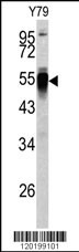
Western blot analysis of PDIA6 antibody (Center K159) (Cat.# AP6662b) in Y79 cell line lysates (35ug/lane). PDIA6 (arrow) was detected using the purified Pab.Application
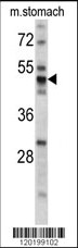
Western blot analysis of PDIA6 antibody (Center K159) (Cat.# AP6662b) in mouse stomach tissue lysates (35ug/lane). PDIA6 (arrow) was detected using the purified Pab.Application
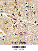
Formalin-fixed and paraffin-embedded human brain tissue reacted with PDIA6 Antibody (Center K159), which was peroxidase-conjugated to the secondary antibody, followed by DAB staining. This data demonstrates the use of this antibody for immunohistochemistry; clinical relevance has not been evaluated.Application
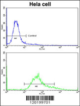
Flow cytometric analysis of hela cells using PDIA6 Antibody (Center K159)(bottom histogram) compared to a negative control cell (top histogram)FITC-conjugated goat-anti-rabbit secondary antibodies were used for the analysis.| Product Name | PDIA6 Antibody (Center K159) |
|---|---|
| Antibody Type | Primary Antibodies |
| Antigen Alias | PDIA6; ERP5; P5; TXNDC7; Protein disulfide-isomerase A6; Endoplasmic reticulum protein 5; Protein disulfide isomerase P5; Thioredoxin domain-containing protein 7 |
| Clonality | Polyclonal |
|---|---|
| Isotype | Ig |
| Host Species | Rabbit |
| Tested Applications | WBIHCFC |
| WB:1:50~100 IHC |
|
| Species Reactivity | Human |
| Concentration | 1mg/ml |
| Gene Synonyms | ERP5 P5 TXNDC7 |
|---|---|
| Alternative Names | PDIA6 ERP5 P5 TXNDC7 Protein disulfide-isomerase A6 Endoplasmic reticulum protein 5 Protein disulfide isomerase P5 Thioredoxin domain-containing protein 7 |
| Molecular Weight(MW) | 48121 Da |
| Function | May function as a chaperone that inhibits aggregation of misfolded proteins. Plays a role in platelet aggregation and activation by agonists such as convulxin, collagen and thrombin |
| Tissue Specificity | Expressed in platelets (at protein level). |
| Cellular Localization | Endoplasmic reticulum lumen (By similarity). Cell membrane. Melanosome. Note=Identified by mass spectrometry in melanosome fractions from stage I to stage IV |
| Entrez Gene | 10130 |
|---|
Application
Western blot analysis of PDIA6 antibody (Center K159) (Cat.# AP6662b) in Y79 cell line lysates (35ug/lane). PDIA6 (arrow) was detected using the purified Pab.
Application
Western blot analysis of PDIA6 antibody (Center K159) (Cat.# AP6662b) in mouse stomach tissue lysates (35ug/lane). PDIA6 (arrow) was detected using the purified Pab.
Application
Formalin-fixed and paraffin-embedded human brain tissue reacted with PDIA6 Antibody (Center K159), which was peroxidase-conjugated to the secondary antibody, followed by DAB staining. This data demonstrates the use of this antibody for immunohistochemistry; clinical relevance has not been evaluated.
Application
Flow cytometric analysis of hela cells using PDIA6 Antibody (Center K159)(bottom histogram) compared to a negative control cell (top histogram)FITC-conjugated goat-anti-rabbit secondary antibodies were used for the analysis.| Application Notes | WB:1:50~100 IHC |
|---|
| Form | Liquid |
|---|---|
| Storage Instructions | For short-term storage, store at 4° C. For long-term storage, aliquot and store at -20ºC or below. Avoid multiple freeze-thaw cycles. |
| Storage Buffer | Purified polyclonal antibody supplied in PBS with 0.09% (W/V) sodium azide. This antibody is prepared by Saturated Ammonium Sulfate (SAS) precipitation followed by dialysis against PBS. |


