Application
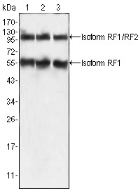
Figure 1: Western blot analysis using anti-PEG10 mAb against HepG2 (1), SMMC-7721 (2) and A549 (3) cell lysate.Application
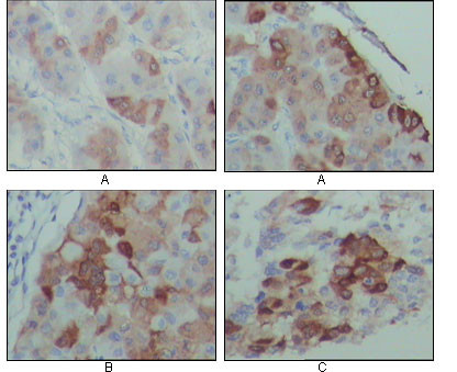
Figure 2: Immunohistochemical analysis of paraffin-embedded human hepatocarcinoma (A), breast carcinoma (B) and lung cancer tissues (C), showing cytoplasmic localization with DAB staining using anti-PEG10 mAb.| Product Name | PEG10 Antibody |
|---|---|
| Antibody Type | Primary Antibodies |
| Antigen Alias | Edr; HB-1; Mar2; MEF3L; Mart2; RGAG3 |
| Clonality | Monoclonal |
|---|---|
| Isotype | IgG1 |
| Clone Number | 4C10A7 |
| Host Species | Mouse |
| Tested Applications | WBIHC |
| WB:1:500 - 2000. IHC |
|
| Species Reactivity | Human |
| Concentration | 1mg/ml |
| Gene Synonyms | EDR KIAA1051 MAR2 MART2 MEF3L1 RGAG |
|---|---|
| Alternative Names | Edr HB-1 Mar2 MEF3L Mart2 RGAG3 |
| Molecular Weight(MW) | 80173 Da |
| Function | Prevents apoptosis in hepatocellular carcinoma (HCC) cells through interaction with SIAH1, a mediator of apoptosis. May also have a role in cell growth promotion and hepatoma formation Inhibits the TGF-beta signaling by interacting with the TGF-beta receptor ALK1. When overexpressed, induces the formation of cellular extension, such as filipodia in association with ALK1 Involved at the immediate early stage of adipocyte differentiation (By similarity). May bind to the 5'-GCCTGTCTTT-3' DNA sequence of the MB1 domain in the myelin basic protein (MBP) promoter (By similarity) |
| Tissue Specificity | Expressed in the cytotrophoblast layer but not in the overlying syncytiotrophoblast of the placenta. Expressed in prostate and breast carcinomas but not in normal breast and prostate epithelial cells. Expressed in the Hep-G2 cell line (at protein level). Expressed in brain, liver, spleen, kidney, thymus, lung, ovary, testis, reactive lymph node, skeletal muscle, adipose tissue and placenta. Expressed in pancreatic and hepatocellular carcinomas (HCC) |
| Cellular Localization | Nucleus. Cytoplasm. Note=Detected predominantly in the cytoplasm of breast and prostate carcinomas, in hepatocellular carcinoma (HCC) and B-cell chronic lymphocytic leukemia (B-CLL) cells and in the Hep-G2 cell line. Colocalized with ALK1 |
| Entrez Gene | 23089 |
|---|---|
| Nucleotide Accession | NP_055883.2 |
Application
Figure 1: Western blot analysis using anti-PEG10 mAb against HepG2 (1), SMMC-7721 (2) and A549 (3) cell lysate.
Application
Figure 2: Immunohistochemical analysis of paraffin-embedded human hepatocarcinoma (A), breast carcinoma (B) and lung cancer tissues (C), showing cytoplasmic localization with DAB staining using anti-PEG10 mAb.| Application Notes | WB:1:500 - 2000. IHC |
|---|
| Form | Liquid |
|---|---|
| Storage Instructions | For short-term storage, store at 4° C. For long-term storage, aliquot and store at -20ºC or below. Avoid multiple freeze-thaw cycles. |
| Storage Buffer | Purified recombinant fragment of human PEG10 expressed in E. Coli. |


