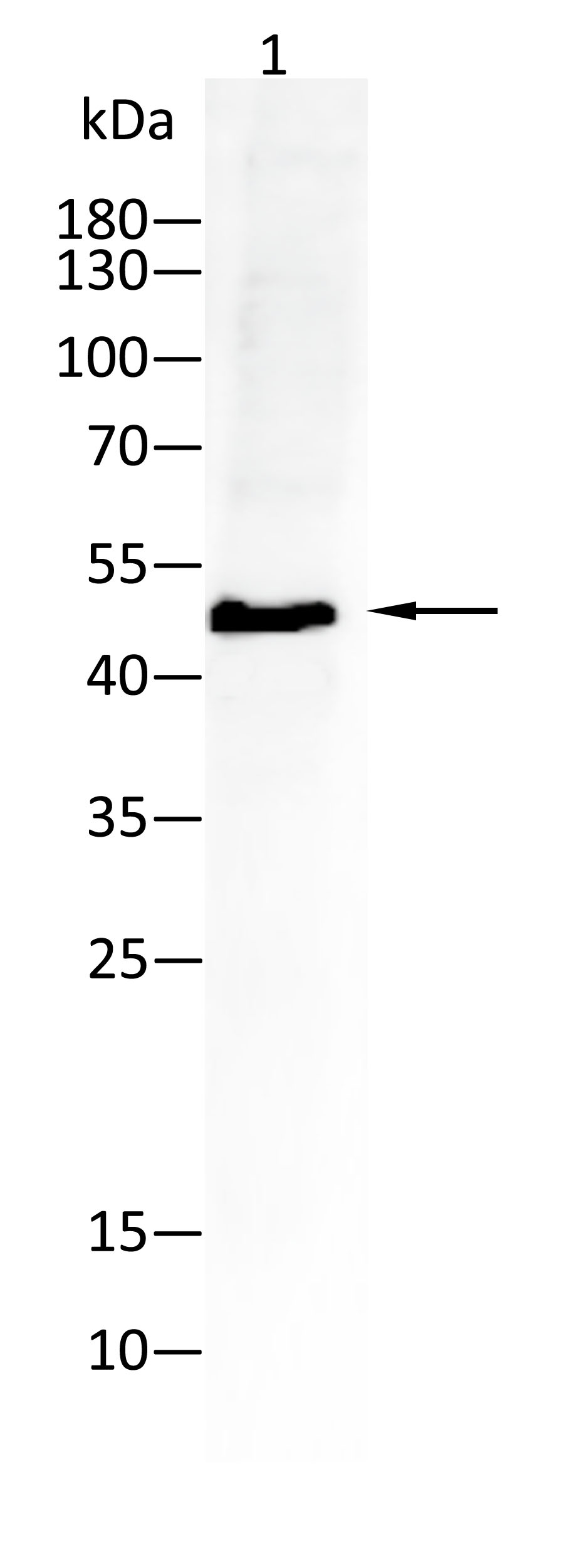
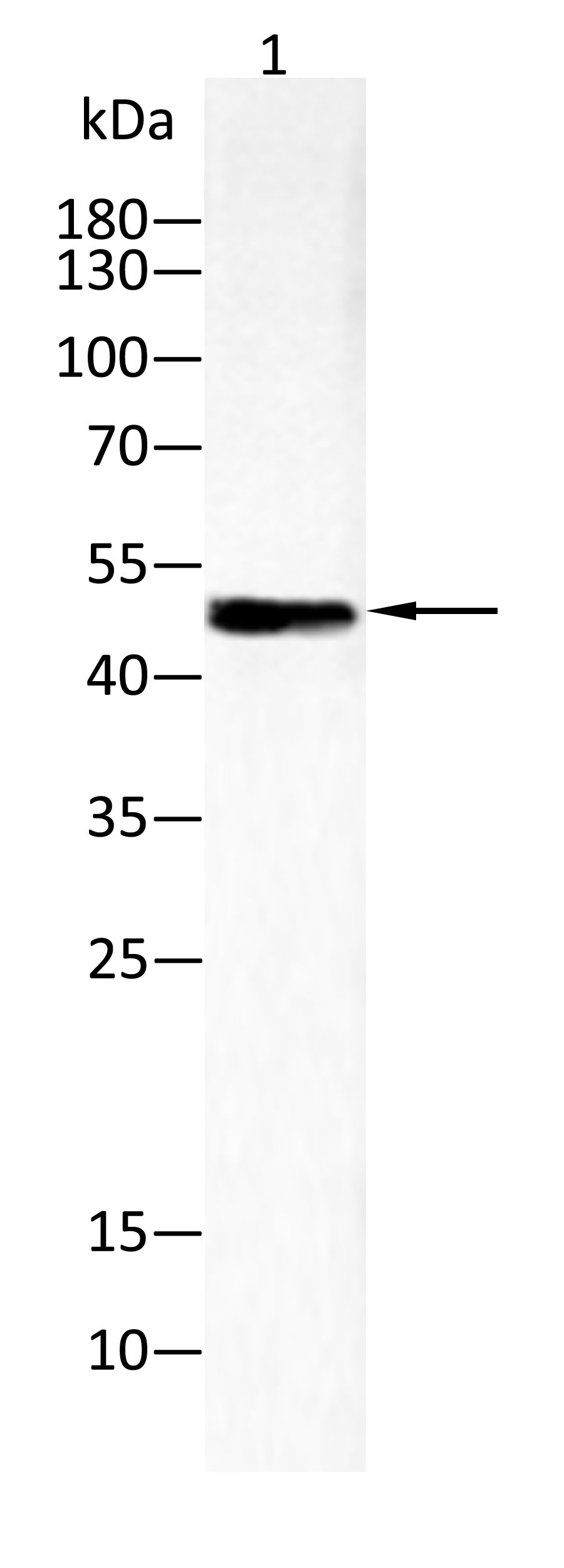

WB
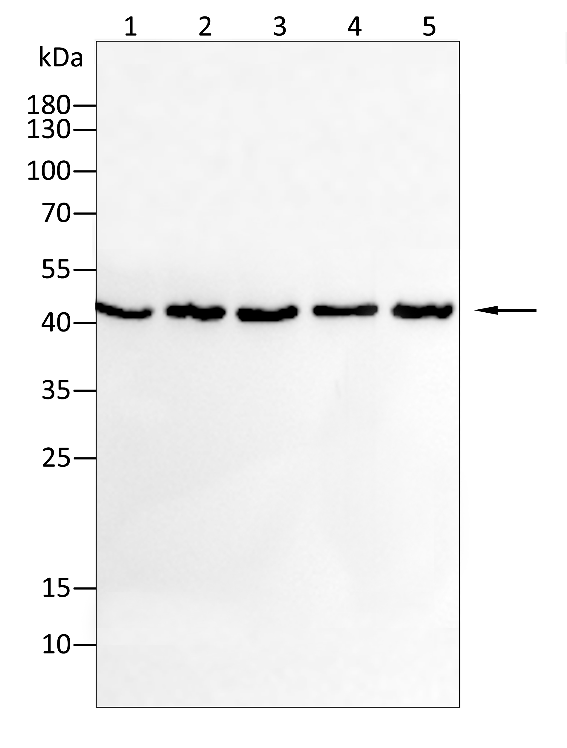
Western blot analysis using Beta-Actin antibody against A431 (1), A549 (2), COS7 (3), Hep G2 (4) and HEK293 (5) cell lysate.12% SDS-PAGE gel.Sample loading: 20μg /lane. Transfer the proteins onto a PVDF membrane (OM790003), and block it with TBST (OM750016) plus skimmed milk powder for one hour. Dilute the primary antibody with the antibody diluent (OM750012) at a ratio of 1:1000, and incubate it overnight at 4°C. Wash the membrane three times with TBST (OM750016), 5 minutes each time. At room temperature, dilute the secondary antibody, Goat Anti-Mouse IgG (H&L) - HRP(OM644366), at a ratio of 1:20000 and incubate for one hour. Wash the membrane three times with TBST (OM750016) again, 5 minutes each time. Use ECL (OM625701) for luminescence.staining time: 60SWB
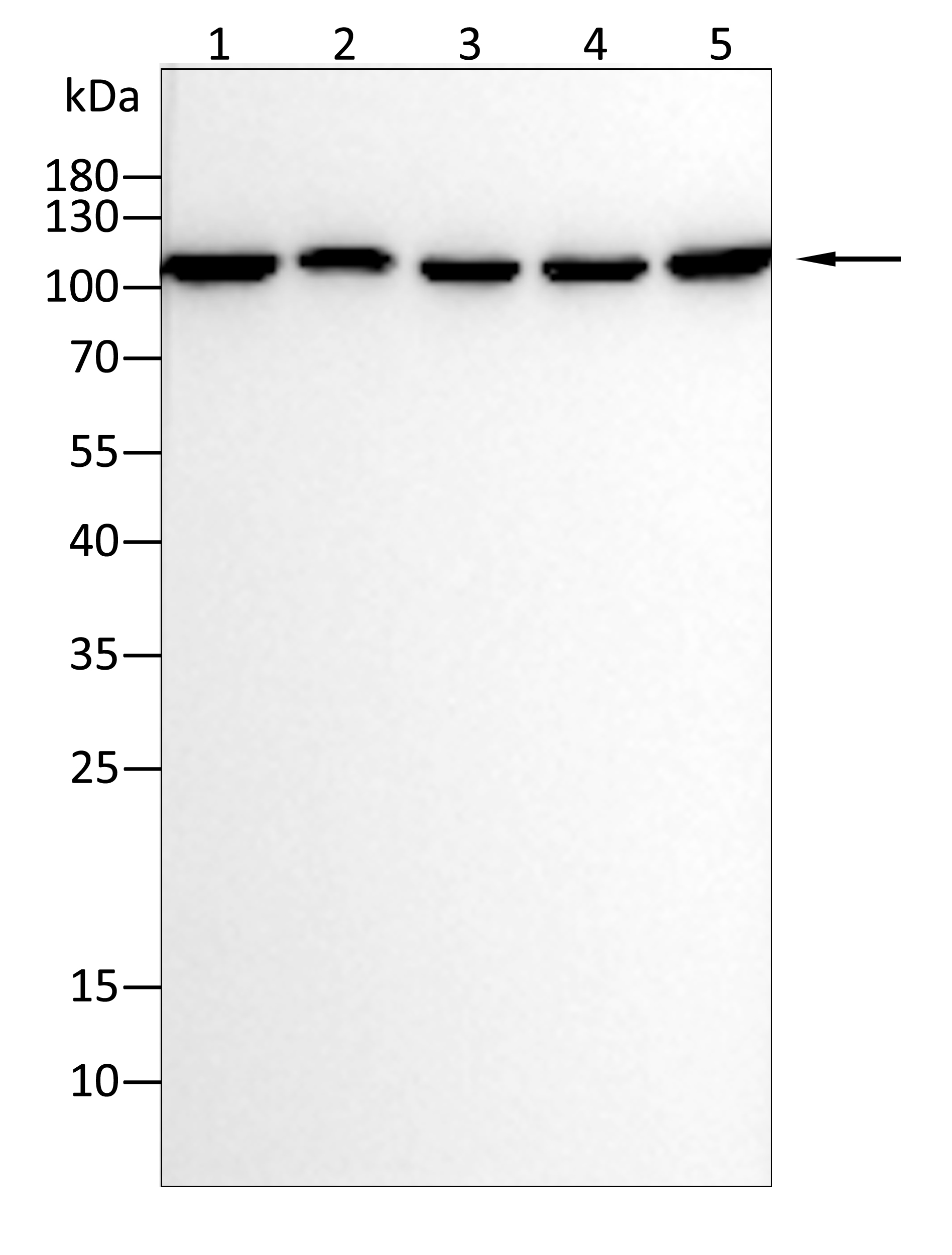
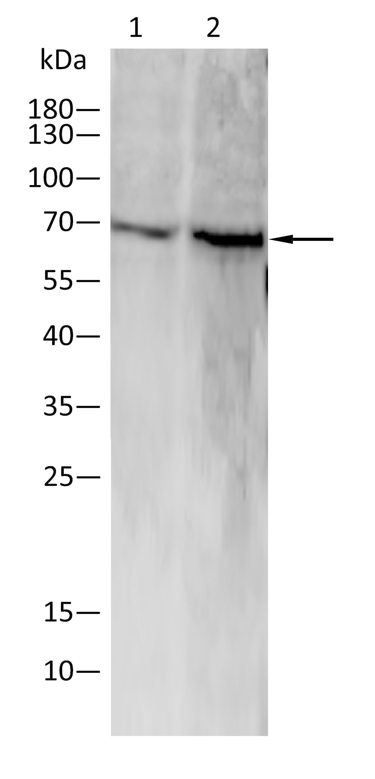
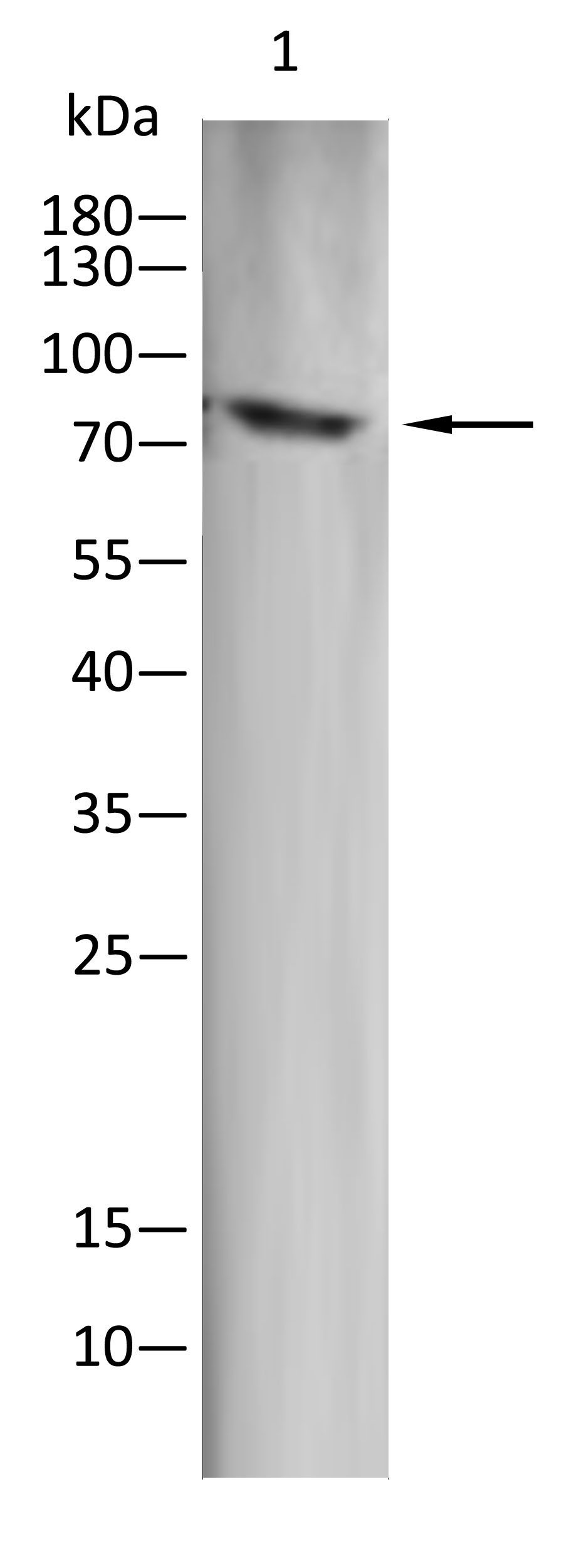
Western blot analysis using LSD1 antibody against A431 (1), A549 (2), Hela (3), Hep G2 (4) and HEK293 (5) cell lysate.12% SDS-PAGE gel.Sample loading: 20μg /lane. Transfer the proteins onto a PVDF membrane (OM790003), and block it with TBST (OM750016) plus skimmed milk powder for one hour. Dilute the primary antibody with the antibody diluent (OM750012) at a ratio of 1:1000, and incubate it overnight at 4°C. Wash the membrane three times with TBST (OM750016), 5 minutes each time. At room temperature, dilute the secondary antibody, Goat Anti-Mouse IgG (H&L) - HRP(OM644366), at a ratio of 1:20000 and incubate for one hour. Wash the membrane three times with TBST (OM750016) again, 5 minutes each time. Use ECL (OM625701) for luminescence.staining time: 60S.IHC
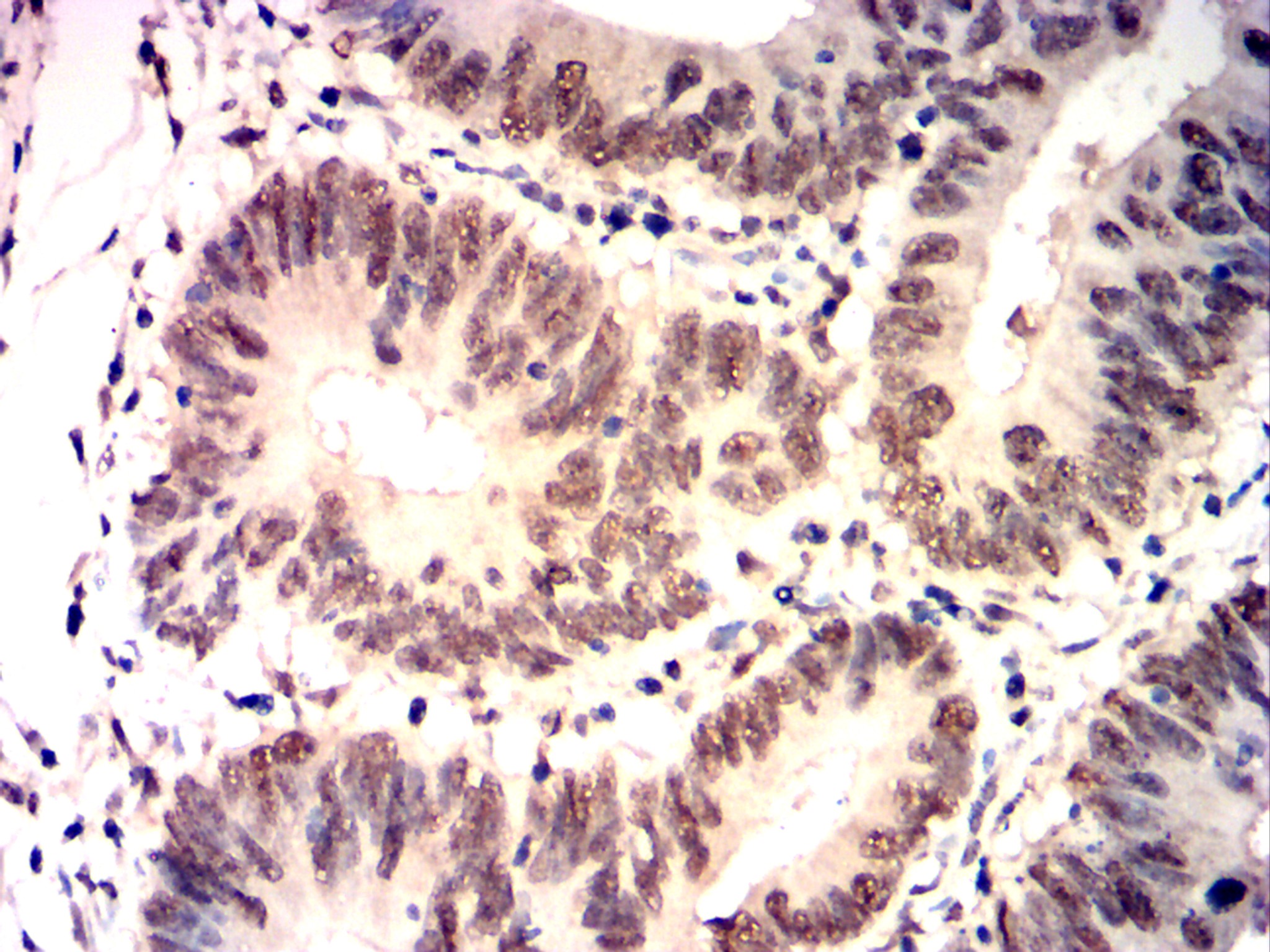
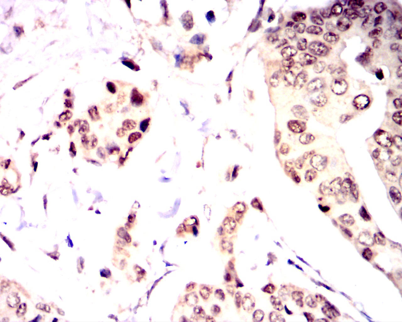
Immunohistochemical analysis of paraffin-embedded rectal cancer tissues using LSD1 antibody with DAB staining.Pre-treat the sections with heat-mediated antigen retrieval using sodium citrate buffer (pH 6.0) (OM750020) for 2 minutes. Wash the sections with ddH₂O and PBS (OM750003). Block the tissue with 10% non-immune goat serum(OM760028) at room temperature for 30 minutes. Incubate the tissue with the primary antibody diluted at a ratio of 1:1500 at 4°C overnight. At room temperature, dilute the secondary antibody, Goat Anti-Mouse IgG (H&L) - HRP(OM644366), at a ratio of 1:200 and incubate for one hour. Use DAB(OM760029)as the chromogenic agent. Counterstain the tissue with hematoxylin, and mount the tissue sections with neutral gum.WB
Western blot analysis using Beta-Actin antibody against A431 (1), A549 (2), COS7 (3), Hep G2 (4) and HEK293 (5) cell lysate.12% SDS-PAGE gel.Sample loading: 20μg /lane. Transfer the proteins onto a PVDF membrane (OM790003), and block it with TBST (OM750016) plus skimmed milk powder for one hour. Dilute the primary antibody with the antibody diluent (OM750012) at a ratio of 1:1000, and incubate it overnight at 4°C. Wash the membrane three times with TBST (OM750016), 5 minutes each time. At room temperature, dilute the secondary antibody, Goat Anti-Mouse IgG (H&L) - HRP(OM644366), at a ratio of 1:20000 and incubate for one hour. Wash the membrane three times with TBST (OM750016) again, 5 minutes each time. Use ECL (OM625701) for luminescence.staining time: 60SWB
Western blot analysis using LSD1 antibody against A431 (1), A549 (2), Hela (3), Hep G2 (4) and HEK293 (5) cell lysate.12% SDS-PAGE gel.Sample loading: 20μg /lane. Transfer the proteins onto a PVDF membrane (OM790003), and block it with TBST (OM750016) plus skimmed milk powder for one hour. Dilute the primary antibody with the antibody diluent (OM750012) at a ratio of 1:1000, and incubate it overnight at 4°C. Wash the membrane three times with TBST (OM750016), 5 minutes each time. At room temperature, dilute the secondary antibody, Goat Anti-Mouse IgG (H&L) - HRP(OM644366), at a ratio of 1:20000 and incubate for one hour. Wash the membrane three times with TBST (OM750016) again, 5 minutes each time. Use ECL (OM625701) for luminescence.staining time: 60S.WB
Western blot analysis using LSD1 antibody against A431 (1), A549 (2), Hela (3), Hep G2 (4) and HEK293 (5) cell lysate.12% SDS-PAGE gel.Sample loading: 20μg /lane. Transfer the proteins onto a PVDF membrane (OM790003), and block it with TBST (OM750016) plus skimmed milk powder for one hour. Dilute the primary antibody with the antibody diluent (OM750012) at a ratio of 1:1000, and incubate it overnight at 4°C. Wash the membrane three times with TBST (OM750016), 5 minutes each time. At room temperature, dilute the secondary antibody, Goat Anti-Mouse IgG (H&L) - HRP(OM644366), at a ratio of 1:20000 and incubate for one hour. Wash the membrane three times with TBST (OM750016) again, 5 minutes each time. Use ECL (OM625701) for luminescence.staining time: 60S.WB
Western blot analysis using Beta-Actin antibody against A431 (1), A549 (2), COS7 (3), Hep G2 (4) and HEK293 (5) cell lysate.12% SDS-PAGE gel.Sample loading: 20μg /lane. Transfer the proteins onto a PVDF membrane (OM790003), and block it with TBST (OM750016) plus skimmed milk powder for one hour. Dilute the primary antibody with the antibody diluent (OM750012) at a ratio of 1:1000, and incubate it overnight at 4°C. Wash the membrane three times with TBST (OM750016), 5 minutes each time. At room temperature, dilute the secondary antibody, Goat Anti-Mouse IgG (H&L) - HRP(OM644366), at a ratio of 1:20000 and incubate for one hour. Wash the membrane three times with TBST (OM750016) again, 5 minutes each time. Use ECL (OM625701) for luminescence.staining time: 60SWB
Western blot analysis using LSD1 antibody against A431 (1), A549 (2), Hela (3), Hep G2 (4) and HEK293 (5) cell lysate.12% SDS-PAGE gel.Sample loading: 20μg /lane. Transfer the proteins onto a PVDF membrane (OM790003), and block it with TBST (OM750016) plus skimmed milk powder for one hour. Dilute the primary antibody with the antibody diluent (OM750012) at a ratio of 1:1000, and incubate it overnight at 4°C. Wash the membrane three times with TBST (OM750016), 5 minutes each time. At room temperature, dilute the secondary antibody, Goat Anti-Mouse IgG (H&L) - HRP(OM644366), at a ratio of 1:20000 and incubate for one hour. Wash the membrane three times with TBST (OM750016) again, 5 minutes each time. Use ECL (OM625701) for luminescence.staining time: 60S.IHC
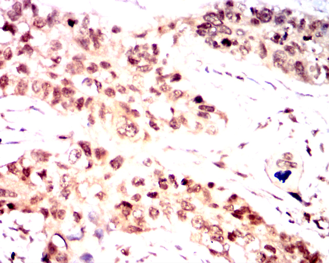
Immunohistochemical analysis of paraffin-embedded breast cancer tissues using OTX2 antibody with DAB staining.Pre-treat the sections with heat-mediated antigen retrieval using sodium citrate buffer (pH 6.0) (OM750020) for 2 minutes. Wash the sections with ddH₂O and PBS (OM750003). Block the tissue with 10% non-immune goat serum(OM760028) at room temperature for 30 minutes. Incubate the tissue with the primary antibody diluted at a ratio of 1:1500 at 4°C overnight. At room temperature, dilute the secondary antibody, Goat Anti-Mouse IgG (H&L) - HRP(OM644366), at a ratio of 1:200 and incubate for one hour. Use DAB(OM760029)as the chromogenic agent. Counterstain the tissue with hematoxylin, and mount the tissue sections with neutral gum.IHC
Immunohistochemical analysis of paraffin-embedded lung cancer tissues using OTX2 antibody with DAB staining.Pre-treat the sections with heat-mediated antigen retrieval using sodium citrate buffer (pH 6.0) (OM750020) for 2 minutes. Wash the sections with ddH₂O and PBS (OM750003). Block the tissue with 10% non-immune goat serum(OM760028) at room temperature for 30 minutes. Incubate the tissue with the primary antibody diluted at a ratio of 1:1500 at 4°C overnight. At room temperature, dilute the secondary antibody, Goat Anti-Mouse IgG (H&L) - HRP(OM644366), at a ratio of 1:200 and incubate for one hour. Use DAB(OM760029)as the chromogenic agent. Counterstain the tissue with hematoxylin, and mount the tissue sections with neutral gum.IHC
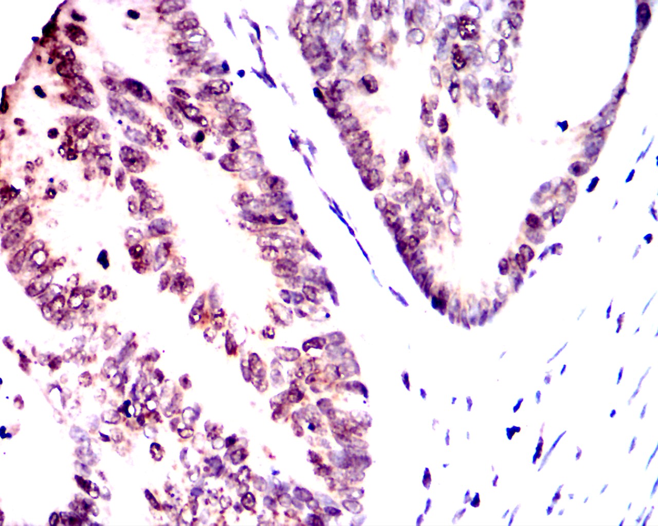
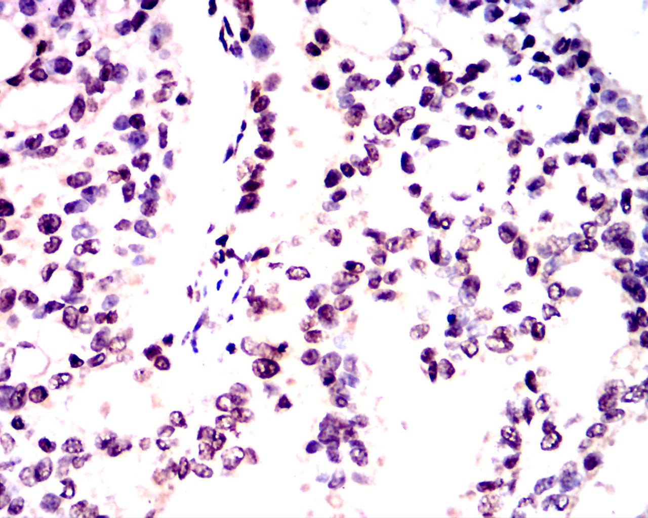
Immunohistochemical analysis of paraffin-embedded colon cancer tissues using Nanog antibody with DAB staining.Pre-treat the sections with heat-mediated antigen retrieval using sodium citrate buffer (pH 6.0) (OM750020) for 2 minutes. Wash the sections with ddH₂O and PBS (OM750003). Block the tissue with 10% non-immune goat serum(OM760028) at room temperature for 30 minutes. Incubate the tissue with the primary antibody diluted at a ratio of 1:1500 at 4°C overnight. At room temperature, dilute the secondary antibody, Goat Anti-Mouse IgG (H&L) - HRP(OM644366), at a ratio of 1:200 and incubate for one hour. Use DAB(OM760029)as the chromogenic agent. Counterstain the tissue with hematoxylin, and mount the tissue sections with neutral gum.IHC
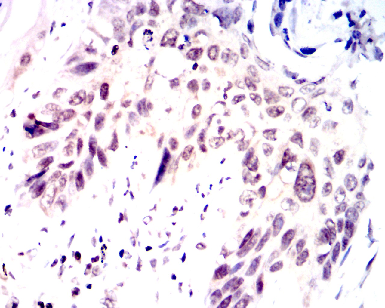
Immunohistochemical analysis of paraffin-embedded lung cancer tissues using Nanog antibody with DAB staining.Pre-treat the sections with heat-mediated antigen retrieval using sodium citrate buffer (pH 6.0) (OM750020) for 2 minutes. Wash the sections with ddH₂O and PBS (OM750003). Block the tissue with 10% non-immune goat serum(OM760028) at room temperature for 30 minutes. Incubate the tissue with the primary antibody diluted at a ratio of 1:1500 at 4°C overnight. At room temperature, dilute the secondary antibody, Goat Anti-Mouse IgG (H&L) - HRP(OM644366), at a ratio of 1:200 and incubate for one hour. Use DAB(OM760029)as the chromogenic agent. Counterstain the tissue with hematoxylin, and mount the tissue sections with neutral gum.IHC
Immunohistochemical analysis of paraffin-embedded lung cancer tissues using Nanog antibody with DAB staining.Pre-treat the sections with heat-mediated antigen retrieval using sodium citrate buffer (pH 6.0) (OM750020) for 2 minutes. Wash the sections with ddH₂O and PBS (OM750003). Block the tissue with 10% non-immune goat serum(OM760028) at room temperature for 30 minutes. Incubate the tissue with the primary antibody diluted at a ratio of 1:1500 at 4°C overnight. At room temperature, dilute the secondary antibody, Goat Anti-Mouse IgG (H&L) - HRP(OM644366), at a ratio of 1:200 and incubate for one hour. Use DAB(OM760029)as the chromogenic agent. Counterstain the tissue with hematoxylin, and mount the tissue sections with neutral gum.IHC
Immunohistochemical analysis of paraffin-embedded thyroid carcinoma tissues using NeuN antibody with DAB staining.Pre-treat the sections with heat-mediated antigen retrieval using sodium citrate buffer (pH 6.0) (OM750020) for 2 minutes. Wash the sections with ddH₂O and PBS (OM750003). Block the tissue with 10% non-immune goat serum(OM760028) at room temperature for 30 minutes. Incubate the tissue with the primary antibody diluted at a ratio of 1:1500 at 4°C overnight. At room temperature, dilute the secondary antibody, Goat Anti-Mouse IgG (H&L) - HRP(OM644366), at a ratio of 1:200 and incubate for one hour. Use DAB(OM760029)as the chromogenic agent. Counterstain the tissue with hematoxylin, and mount the tissue sections with neutral gum.| Product Name | Goat Anti-Mouse IgG (H&L) - HRP |
|---|---|
| Antibody Type | Secondary Antibodies |
| Immunogen | Mouse IgG |
| Conjugation | HRP |
| Clonality | Polyclonal |
|---|---|
| Host Species | Goat |
| Tested Applications | ICCIHCWB |
| WB:1:5000-1:20000 IHC:1:500-1:1000 ICC:1:500-1:1000 |
|
| Species Reactivity | Mouse |
| Concentration | 1mg/ml |
| Purification | Affinity purified |
WB
Western blot analysis using Beta-Actin antibody against A431 (1), A549 (2), COS7 (3), Hep G2 (4) and HEK293 (5) cell lysate.12% SDS-PAGE gel.Sample loading: 20μg /lane. Transfer the proteins onto a PVDF membrane (OM790003), and block it with TBST (OM750016) plus skimmed milk powder for one hour. Dilute the primary antibody with the antibody diluent (OM750012) at a ratio of 1:1000, and incubate it overnight at 4°C. Wash the membrane three times with TBST (OM750016), 5 minutes each time. At room temperature, dilute the secondary antibody, Goat Anti-Mouse IgG (H&L) - HRP(OM644366), at a ratio of 1:20000 and incubate for one hour. Wash the membrane three times with TBST (OM750016) again, 5 minutes each time. Use ECL (OM625701) for luminescence.staining time: 60S
WB
Western blot analysis using LSD1 antibody against A431 (1), A549 (2), Hela (3), Hep G2 (4) and HEK293 (5) cell lysate.12% SDS-PAGE gel.Sample loading: 20μg /lane. Transfer the proteins onto a PVDF membrane (OM790003), and block it with TBST (OM750016) plus skimmed milk powder for one hour. Dilute the primary antibody with the antibody diluent (OM750012) at a ratio of 1:1000, and incubate it overnight at 4°C. Wash the membrane three times with TBST (OM750016), 5 minutes each time. At room temperature, dilute the secondary antibody, Goat Anti-Mouse IgG (H&L) - HRP(OM644366), at a ratio of 1:20000 and incubate for one hour. Wash the membrane three times with TBST (OM750016) again, 5 minutes each time. Use ECL (OM625701) for luminescence.staining time: 60S.
IHC
Immunohistochemical analysis of paraffin-embedded rectal cancer tissues using LSD1 antibody with DAB staining.Pre-treat the sections with heat-mediated antigen retrieval using sodium citrate buffer (pH 6.0) (OM750020) for 2 minutes. Wash the sections with ddH₂O and PBS (OM750003). Block the tissue with 10% non-immune goat serum(OM760028) at room temperature for 30 minutes. Incubate the tissue with the primary antibody diluted at a ratio of 1:1500 at 4°C overnight. At room temperature, dilute the secondary antibody, Goat Anti-Mouse IgG (H&L) - HRP(OM644366), at a ratio of 1:200 and incubate for one hour. Use DAB(OM760029)as the chromogenic agent. Counterstain the tissue with hematoxylin, and mount the tissue sections with neutral gum.
WB
Western blot analysis using Beta-Actin antibody against A431 (1), A549 (2), COS7 (3), Hep G2 (4) and HEK293 (5) cell lysate.12% SDS-PAGE gel.Sample loading: 20μg /lane. Transfer the proteins onto a PVDF membrane (OM790003), and block it with TBST (OM750016) plus skimmed milk powder for one hour. Dilute the primary antibody with the antibody diluent (OM750012) at a ratio of 1:1000, and incubate it overnight at 4°C. Wash the membrane three times with TBST (OM750016), 5 minutes each time. At room temperature, dilute the secondary antibody, Goat Anti-Mouse IgG (H&L) - HRP(OM644366), at a ratio of 1:20000 and incubate for one hour. Wash the membrane three times with TBST (OM750016) again, 5 minutes each time. Use ECL (OM625701) for luminescence.staining time: 60S
WB
Western blot analysis using LSD1 antibody against A431 (1), A549 (2), Hela (3), Hep G2 (4) and HEK293 (5) cell lysate.12% SDS-PAGE gel.Sample loading: 20μg /lane. Transfer the proteins onto a PVDF membrane (OM790003), and block it with TBST (OM750016) plus skimmed milk powder for one hour. Dilute the primary antibody with the antibody diluent (OM750012) at a ratio of 1:1000, and incubate it overnight at 4°C. Wash the membrane three times with TBST (OM750016), 5 minutes each time. At room temperature, dilute the secondary antibody, Goat Anti-Mouse IgG (H&L) - HRP(OM644366), at a ratio of 1:20000 and incubate for one hour. Wash the membrane three times with TBST (OM750016) again, 5 minutes each time. Use ECL (OM625701) for luminescence.staining time: 60S.
WB
Western blot analysis using Beta-Actin antibody against A431 (1), A549 (2), COS7 (3), Hep G2 (4) and HEK293 (5) cell lysate.12% SDS-PAGE gel.Sample loading: 20μg /lane. Transfer the proteins onto a PVDF membrane (OM790003), and block it with TBST (OM750016) plus skimmed milk powder for one hour. Dilute the primary antibody with the antibody diluent (OM750012) at a ratio of 1:1000, and incubate it overnight at 4°C. Wash the membrane three times with TBST (OM750016), 5 minutes each time. At room temperature, dilute the secondary antibody, Goat Anti-Mouse IgG (H&L) - HRP(OM644366), at a ratio of 1:20000 and incubate for one hour. Wash the membrane three times with TBST (OM750016) again, 5 minutes each time. Use ECL (OM625701) for luminescence.staining time: 60S
WB
Western blot analysis using LSD1 antibody against A431 (1), A549 (2), Hela (3), Hep G2 (4) and HEK293 (5) cell lysate.12% SDS-PAGE gel.Sample loading: 20μg /lane. Transfer the proteins onto a PVDF membrane (OM790003), and block it with TBST (OM750016) plus skimmed milk powder for one hour. Dilute the primary antibody with the antibody diluent (OM750012) at a ratio of 1:1000, and incubate it overnight at 4°C. Wash the membrane three times with TBST (OM750016), 5 minutes each time. At room temperature, dilute the secondary antibody, Goat Anti-Mouse IgG (H&L) - HRP(OM644366), at a ratio of 1:20000 and incubate for one hour. Wash the membrane three times with TBST (OM750016) again, 5 minutes each time. Use ECL (OM625701) for luminescence.staining time: 60S.
IHC
Immunohistochemical analysis of paraffin-embedded breast cancer tissues using OTX2 antibody with DAB staining.Pre-treat the sections with heat-mediated antigen retrieval using sodium citrate buffer (pH 6.0) (OM750020) for 2 minutes. Wash the sections with ddH₂O and PBS (OM750003). Block the tissue with 10% non-immune goat serum(OM760028) at room temperature for 30 minutes. Incubate the tissue with the primary antibody diluted at a ratio of 1:1500 at 4°C overnight. At room temperature, dilute the secondary antibody, Goat Anti-Mouse IgG (H&L) - HRP(OM644366), at a ratio of 1:200 and incubate for one hour. Use DAB(OM760029)as the chromogenic agent. Counterstain the tissue with hematoxylin, and mount the tissue sections with neutral gum.
IHC
Immunohistochemical analysis of paraffin-embedded lung cancer tissues using OTX2 antibody with DAB staining.Pre-treat the sections with heat-mediated antigen retrieval using sodium citrate buffer (pH 6.0) (OM750020) for 2 minutes. Wash the sections with ddH₂O and PBS (OM750003). Block the tissue with 10% non-immune goat serum(OM760028) at room temperature for 30 minutes. Incubate the tissue with the primary antibody diluted at a ratio of 1:1500 at 4°C overnight. At room temperature, dilute the secondary antibody, Goat Anti-Mouse IgG (H&L) - HRP(OM644366), at a ratio of 1:200 and incubate for one hour. Use DAB(OM760029)as the chromogenic agent. Counterstain the tissue with hematoxylin, and mount the tissue sections with neutral gum.
IHC
Immunohistochemical analysis of paraffin-embedded colon cancer tissues using Nanog antibody with DAB staining.Pre-treat the sections with heat-mediated antigen retrieval using sodium citrate buffer (pH 6.0) (OM750020) for 2 minutes. Wash the sections with ddH₂O and PBS (OM750003). Block the tissue with 10% non-immune goat serum(OM760028) at room temperature for 30 minutes. Incubate the tissue with the primary antibody diluted at a ratio of 1:1500 at 4°C overnight. At room temperature, dilute the secondary antibody, Goat Anti-Mouse IgG (H&L) - HRP(OM644366), at a ratio of 1:200 and incubate for one hour. Use DAB(OM760029)as the chromogenic agent. Counterstain the tissue with hematoxylin, and mount the tissue sections with neutral gum.
IHC
Immunohistochemical analysis of paraffin-embedded lung cancer tissues using Nanog antibody with DAB staining.Pre-treat the sections with heat-mediated antigen retrieval using sodium citrate buffer (pH 6.0) (OM750020) for 2 minutes. Wash the sections with ddH₂O and PBS (OM750003). Block the tissue with 10% non-immune goat serum(OM760028) at room temperature for 30 minutes. Incubate the tissue with the primary antibody diluted at a ratio of 1:1500 at 4°C overnight. At room temperature, dilute the secondary antibody, Goat Anti-Mouse IgG (H&L) - HRP(OM644366), at a ratio of 1:200 and incubate for one hour. Use DAB(OM760029)as the chromogenic agent. Counterstain the tissue with hematoxylin, and mount the tissue sections with neutral gum.
IHC
Immunohistochemical analysis of paraffin-embedded thyroid carcinoma tissues using NeuN antibody with DAB staining.Pre-treat the sections with heat-mediated antigen retrieval using sodium citrate buffer (pH 6.0) (OM750020) for 2 minutes. Wash the sections with ddH₂O and PBS (OM750003). Block the tissue with 10% non-immune goat serum(OM760028) at room temperature for 30 minutes. Incubate the tissue with the primary antibody diluted at a ratio of 1:1500 at 4°C overnight. At room temperature, dilute the secondary antibody, Goat Anti-Mouse IgG (H&L) - HRP(OM644366), at a ratio of 1:200 and incubate for one hour. Use DAB(OM760029)as the chromogenic agent. Counterstain the tissue with hematoxylin, and mount the tissue sections with neutral gum.| Application Notes | WB:1:5000-1:20000 IHC:1:500-1:1000 ICC:1:500-1:1000 |
|---|
| Form | Liquid |
|---|---|
| Storage Instructions | Shipped at 4°C. Upon delivery aliquot and store at -20°C for one year. Avoid freeze/thaw cycles. |
| Storage Buffer | Liquid in 0.01M Phosphate Buffered Saline, pH 7.2, containing 1% BSA, 50% glycerol, 0.02% Sodium Azide. |


 Data sheet for OM644366