Application
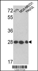
Western blot analysis of PGAM1 Antibody (Center) in Y79, MDA-MB231, HepG2 cell line lysates (35ug/lane). PGAM1 (arrow) was detected using the purified Pab.(2ug/ml)Application
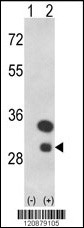
Western blot analysis of PGAM1 (arrow) using rabbit polyclonal PGAM1 Antibody (Center) . 293 cell lysates (2 ug/lane) either nontransfected (Lane 1) or transiently transfected with the PGAM1 gene (Lane 2) .Application
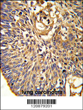
Formalin-fixed and paraffin-embedded human lung carcinoma reacted with PGAM1 Antibody (Center), which was peroxidase-conjugated to the secondary antibody, followed by DAB staining. This data demonstrates the use of this antibody for immunohistochemistry; clinical relevance has not been evaluated.Application
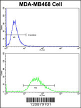
Flow cytometric analysis of MDA-MB468 cells using PGAM1 Antibody (Center)(bottom histogram) compared to a negative control cell (top histogram)FITC-conjugated goat-anti-rabbit secondary antibodies were used for the analysis.| Product Name | PGAM1 Antibody (Center) |
|---|---|
| Antibody Type | Primary Antibodies |
| Antigen Alias | PGAM1; PGAMA; Phosphoglycerate mutase 1; BPG-dependent PGAM 1; Phosphoglycerate mutase isozyme B |
| Clonality | Polyclonal |
|---|---|
| Isotype | Ig |
| Host Species | Rabbit |
| Tested Applications | WBIHCFC |
| WB:1:50~100 IHC |
|
| Species Reactivity | Human |
| Concentration | 1mg/ml |
| Gene Synonyms | PGAMA |
|---|---|
| Alternative Names | PGAM1 PGAMA Phosphoglycerate mutase 1 BPG-dependent PGAM 1 Phosphoglycerate mutase isozyme B |
| Molecular Weight(MW) | 28804 Da |
| Function | Interconversion of 3- and 2-phosphoglycerate with 2,3- bisphosphoglycerate as the primer of the reaction. Can also catalyze the reaction of EC 5.4.2.4 (synthase) and EC 3.1.3.13 (phosphatase), but with a reduced activity |
| Tissue Specificity | In mammalian tissues there are two types of phosphoglycerate mutase isozymes: type-M in muscles and type-B in other tissues |
| Entrez Gene | 5223 |
|---|
Application
Western blot analysis of PGAM1 Antibody (Center) in Y79, MDA-MB231, HepG2 cell line lysates (35ug/lane). PGAM1 (arrow) was detected using the purified Pab.(2ug/ml)
Application
Western blot analysis of PGAM1 (arrow) using rabbit polyclonal PGAM1 Antibody (Center) . 293 cell lysates (2 ug/lane) either nontransfected (Lane 1) or transiently transfected with the PGAM1 gene (Lane 2) .
Application
Formalin-fixed and paraffin-embedded human lung carcinoma reacted with PGAM1 Antibody (Center), which was peroxidase-conjugated to the secondary antibody, followed by DAB staining. This data demonstrates the use of this antibody for immunohistochemistry; clinical relevance has not been evaluated.
Application
Flow cytometric analysis of MDA-MB468 cells using PGAM1 Antibody (Center)(bottom histogram) compared to a negative control cell (top histogram)FITC-conjugated goat-anti-rabbit secondary antibodies were used for the analysis.| Application Notes | WB:1:50~100 IHC |
|---|
| Form | Liquid |
|---|---|
| Storage Instructions | For short-term storage, store at 4° C. For long-term storage, aliquot and store at -20ºC or below. Avoid multiple freeze-thaw cycles. |
| Storage Buffer | Purified polyclonal antibody supplied in PBS with 0.09% (W/V) sodium azide. This antibody is prepared by Saturated Ammonium Sulfate (SAS) precipitation followed by dialysis against PBS. |


