Application
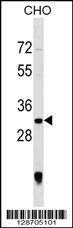
PGAM5 Antibody (C-term) western blot analysis in CHO cell line lysates (35ug/lane).This demonstrates the PGAM5 antibody detected the PGAM5 protein (arrow).| Product Name | PGAM5 Antibody (C-term) |
|---|---|
| Antibody Type | Primary Antibodies |
| Antigen Alias | PGAM5; Serine/threonine-protein phosphatase PGAM5, mitochondrial; Bcl-XL-binding protein v68; Phosphoglycerate mutase family member 5 |
| Clonality | Polyclonal |
|---|---|
| Isotype | Ig |
| Host Species | Rabbit |
| Tested Applications | WB |
| WB:1:100~500 |
|
| Species Reactivity | Human |
| Concentration | 1mg/ml |
| Alternative Names | PGAM5 Serine/threonine-protein phosphatase PGAM5 mitochondrial Bcl-XL-binding protein v68 Phosphoglycerate mutase family member 5 |
|---|---|
| Molecular Weight(MW) | 32004 Da |
| Function | Displays phosphatase activity for serine/threonine residues, and, dephosphorylates and activates MAP3K5 kinase. Has apparently no phosphoglycerate mutase activity. May be regulator of mitochondrial dynamics. Substrate for a KEAP1-dependent ubiquitin ligase complex. Contributes to the repression of NFE2L2- dependent gene expression. Acts as a central mediator for programmed necrosis induced by TNF, by reactive oxygen species and by calcium ionophore |
| Tissue Specificity | This PGAM5 antibody is generated from rabbits immunized with a KLH conjugated synthetic peptide between 196-225 amino acids from the C-terminal region of human PGAM5. |
| Cellular Localization | Mitochondrion outer membrane; Single-pass membrane protein. Note=Isoform 2 overexpression results in the formation of disconnected punctuate mitochondria distributed throughout the cytoplasm. Isoform 1 overexpression results in the clustering of mitochondria around the nucleus |
| Entrez Gene | 192111 |
|---|---|
| Nucleotide Accession | NP_612642.2 |
Application
PGAM5 Antibody (C-term) western blot analysis in CHO cell line lysates (35ug/lane).This demonstrates the PGAM5 antibody detected the PGAM5 protein (arrow).| Application Notes | WB:1:100~500 |
|---|
| Form | Liquid |
|---|---|
| Storage Instructions | For short-term storage, store at 4° C. For long-term storage, aliquot and store at -20ºC or below. Avoid multiple freeze-thaw cycles. |
| Storage Buffer | Purified polyclonal antibody supplied in PBS with 0.09% (W/V) sodium azide. This antibody is purified through a protein A column, followed by peptide affinity purification. |


