Application
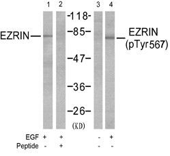
Western blot analysis of extracts from A431 cells, untreated or EGFtreated(200ng/ml, 30min) using Ezrin Antibody (T567) (#AE1012a, Lane 1and 2) and Phospho-Ezrin-T567 Antibody (#AE1012b, Lane 3 and 4).| Product Name | Phospho-Ezrin-T567 Antibody |
|---|---|
| Antibody Type | Primary Antibodies |
| Modification | p-T567 |
| Clonality | Polyclonal |
|---|---|
| Isotype | IgG |
| Host Species | Rabbit |
| Tested Applications | WB |
| WB:1:500~1:1000 |
|
| Species Reactivity | Human |
| Concentration | 1mg/ml |
| Gene Synonyms | VIL2 |
|---|---|
| Alternative Names | Villin-2 Cytovillin p81 Ezrin |
| Molecular Weight(MW) | 69413 Da |
| Function | Probably involved in connections of major cytoskeletal structures to the plasma membrane. In epithelial cells, required for the formation of microvilli and membrane ruffles on the apical pole. Along with PLEKHG6, required for normal macropinocytosis |
| Tissue Specificity | Expressed in cerebral cortex, basal ganglia, hippocampus, hypophysis, and optic nerve. Weakly expressed in brain stem and diencephalon. Stronger expression was detected in gray matter of frontal lobe compared to white matter (at protein level). Component of the microvilli of intestinal epithelial cells. Preferentially expressed in astrocytes of hippocampus, frontal cortex, thalamus, parahippocampal cortex, amygdala, insula, and corpus callosum. Not detected in neurons in most tissues studied |
| Cellular Localization | Apical cell membrane; Peripheral membrane protein; Cytoplasmic side. Cell projection. Cell projection, microvillus membrane; Peripheral membrane protein; Cytoplasmic side. Cell projection, ruffle membrane; Peripheral membrane protein; Cytoplasmic side. Cytoplasm, cell cortex. Cytoplasm, cytoskeleton. Note=Localization to the apical membrane of parietal cells depends on the interaction with MPP5. Localizes to cell extensions and peripheral processes of astrocytes (By similarity) Microvillar peripheral membrane protein (cytoplasmic side) |
| Entrez Gene | 7430 |
|---|
Application
Western blot analysis of extracts from A431 cells, untreated or EGFtreated(200ng/ml, 30min) using Ezrin Antibody (T567) (#AE1012a, Lane 1and 2) and Phospho-Ezrin-T567 Antibody (#AE1012b, Lane 3 and 4).| Application Notes | WB:1:500~1:1000 |
|---|
| Form | Liquid |
|---|---|
| Storage Instructions | For short-term storage, store at 4° C. For long-term storage, aliquot and store at -20ºC or below. Avoid multiple freeze-thaw cycles. |
| Storage Buffer | affinity Purified IgG, in PBS, 0.02% sodium azide and 50% glycerol. |


