Application
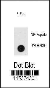
Dot blot analysis of anti-Phospho-GIT1-pY510 Antibody on nitrocellulose membrane. 50ng of Phospho-peptide or Non Phospho-peptide per dot were adsorbed. Antibody working concentrations are 0.5ug per ml.| Product Name | Phospho-GIT1-pY510 Antibody |
|---|---|
| Antibody Type | Primary Antibodies |
| Modification | p-Y510 |
| Clonality | Polyclonal |
|---|---|
| Isotype | Ig |
| Host Species | Rabbit |
| Tested Applications | DB |
| DB:1:500 |
|
| Species Reactivity | Human |
| Concentration | 1mg/ml |
| Alternative Names | GIT1 ARF GTPase-activating protein GIT1 Cool-associated and tyrosine-phosphorylated protein 1 G protein-coupled receptor kinase-interactor 1 GRK-interacting protein 1 |
|---|---|
| Molecular Weight(MW) | 84341 Da |
| Function | GTPase-activating protein for the ADP ribosylation factor family. May serve as a scaffold to bring together molecules to form signaling modules controlling vesicle trafficking, adhesion and cytoskeletal organization. Increases the speed of cell migration, as well as the size and rate of formation of protrusions, possibly by targeting PAK1 to adhesions and the leading edge of lamellipodia. Sequesters inactive non-tyrosine- phosphorylated paxillin in cytoplasmic complexes |
| Tissue Specificity | This Phospho-GIT1-pY510 antibody is generated from rabbits immunized with a KLH conjugated synthetic phosphopeptide between 495~524 amino acids surrounding Y510 of human GIT1. |
| Cellular Localization | Cytoplasm. Note=Cycles between at least 3 distinct intracellular compartments, including focal adhesions, cytoplasmic complexes and membrane protrusions. During cell migration, when cells detach, moves from the adhesions into the cytoplasmic complexes towards the leading edge, while, when cells adhere, it is found in vinculin-containing adhesions. Recruitment to adhesions may be mediated by active tyrosine-phosphorylated paxillin |
| Entrez Gene | 28964 |
|---|
Application
Dot blot analysis of anti-Phospho-GIT1-pY510 Antibody on nitrocellulose membrane. 50ng of Phospho-peptide or Non Phospho-peptide per dot were adsorbed. Antibody working concentrations are 0.5ug per ml.| Application Notes | DB:1:500 |
|---|
| Form | Liquid |
|---|---|
| Storage Instructions | For short-term storage, store at 4° C. For long-term storage, aliquot and store at -20ºC or below. Avoid multiple freeze-thaw cycles. |
| Storage Buffer | Purified polyclonal antibody supplied in PBS with 0.09% (W/V) sodium azide. This antibody is purified through a protein A column, followed by two-step phosphospecific peptide affinity purification. |


