Application
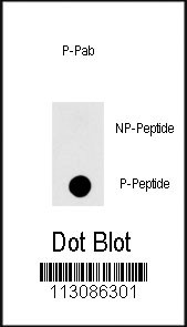
Dot blot analysis of anti-HSPB1-pS83 Phospho-specific Pab on nitrocellulose membrane. 50ng of Phospho-peptide or Non Phospho-peptide per dot were adsorbed. Antibody working concentrations are 0.5ug per ml.| Product Name | Phospho-HSPB1-pS83 Antibody |
|---|---|
| Antibody Type | Primary Antibodies |
| Modification | p-S83 |
| Clonality | Polyclonal |
|---|---|
| Isotype | Ig |
| Host Species | Rabbit |
| Tested Applications | DB |
| DB:1:500 |
|
| Species Reactivity | Human |
| Concentration | 1mg/ml |
| Gene Synonyms | HSP27, HSP28 |
|---|---|
| Alternative Names | HSPB1 HSP27 HSP28 Heat shock protein beta-1 28 kDa heat shock protein Estrogen-regulated 24 kDa protein Heat shock 27 kDa protein Stress-responsive protein 27 |
| Molecular Weight(MW) | 22783 Da |
| Function | Involved in stress resistance and actin organization. |
| Tissue Specificity | Detected in all tissues tested: skeletal muscle, heart, aorta, large intestine, small intestine, stomach, esophagus, bladder, adrenal gland, thyroid, pancreas, testis, adipose tissue, kidney, liver, spleen, cerebral cortex, blood serum and cerebrospinal fluid. Highest levels are found in the heart and in tissues composed of striated and smooth muscle |
| Cellular Localization | Cytoplasm. Nucleus. Cytoplasm, cytoskeleton, spindle. Note=Cytoplasmic in interphase cells. Colocalizes with mitotic spindles in mitotic cells. Translocates to the nucleus during heat shock and resides in sub-nuclear structures known as SC35 speckles or nuclear splicing speckles |
| Entrez Gene | 3315 |
|---|
Application
Dot blot analysis of anti-HSPB1-pS83 Phospho-specific Pab on nitrocellulose membrane. 50ng of Phospho-peptide or Non Phospho-peptide per dot were adsorbed. Antibody working concentrations are 0.5ug per ml.| Application Notes | DB:1:500 |
|---|
| Form | Liquid |
|---|---|
| Storage Instructions | For short-term storage, store at 4° C. For long-term storage, aliquot and store at -20ºC or below. Avoid multiple freeze-thaw cycles. |
| Storage Buffer | Purified polyclonal antibody supplied in PBS with 0.09% (W/V) sodium azide. This antibody is first purified by protein A affinity chromatography. Then, the antibody fraction is peptide affinity purified in a 2-step procedure with peptides. The antibody is eluted with high and low pH buffers and neutralized immediately, followed by dialysis against PBS. |


