Application
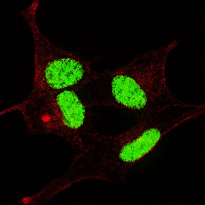
Fluorescent confocal image of SY5Y cells stained with phospho- LIN28- S134 antibody. SY5Y cells were fixed with 4% PFA (20 min), permeabilized with Triton X-100 (0.2%, 30 min). Cells were then incubated with AP3732a phospho- LIN28- S134 primary antibody (1:100, 2 h at room temperature). For secondary antibody, Alexa Fluor® 488 conjugated donkey anti-rabbit antibody (green) was used (1:1000, 1h). Nuclei were counterstained with Hoechst 33342 (blue) (10 μg/ml, 5 min). Note the highly specific localization of the phospho- LIN28 immunosignal mainly to the nucleus, supported by Human Protein Atlas Data (http://www.proteinatlas.org/ENSG00000131914).Application
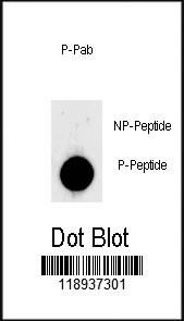
Dot blot analysis of anti-Phospho-LIN28-S134 Phospho-specific Pab on nitrocellulose membrane. 50ng of Phospho-peptide or Non Phospho-peptide per dot were adsorbed. Antibody working concentrations are 0.5ug per ml.| Product Name | Phospho-LIN28- S134 Antibody |
|---|---|
| Antibody Type | Primary Antibodies |
| Modification | p-S134 |
| Clonality | Polyclonal |
|---|---|
| Isotype | Ig |
| Host Species | Rabbit |
| Tested Applications | IFDB |
| IF:1:100 DB |
|
| Species Reactivity | Human |
| Concentration | 1mg/ml |
| Gene Synonyms | CSDD1, LIN28, ZCCHC1 |
|---|---|
| Alternative Names | LIN28A CSDD1 LIN28 ZCCHC1 Protein lin-28 homolog A Zinc finger CCHC domain-containing protein 1 |
| Molecular Weight(MW) | 22743 Da |
| Function | Acts as a 'translational enhancer', driving specific mRNAs to polysomes and thus increasing the efficiency of protein synthesis. Its association with the translational machinery and target mRNAs results in an increased number of initiation events per molecule of mRNA and, indirectly, in stabilizing the mRNAs Binds IGF2 mRNA, MYOD1 mRNA, ARBP/36B4 ribosomal protein mRNA and its own mRNA. Essential for skeletal muscle differentiation program through the translational up-regulation of IGF2 expression (By similarity). Acts as a suppressor of microRNA (miRNA) biogenesis by specifically binding the precursor let-7 (pre-let- 7), a miRNA precursor. Acts by binding pre-let-7 and recruiting ZCCHC11/TUT4 uridylyltransferase, leading to the terminal uridylation of pre-let-7. Uridylated pre-let-7 miRNAs fail to be processed by Dicer and undergo degradation. Degradation of pre- let-7 in embryonic stem (ES) cells contributes to the maintenance of ES cells. In contrast, LIN28A down-regulation in neural stem cells by miR-125, allows the processing of pre-let-7. Specifically recognizes the 5'-GGAG-3' motif in the terminal loop of pre-let-7 Also recognizes and binds non pre-let-7 pre-miRNAs that contain the 5'-GGAG-3' motif in the terminal loop, leading to their terminal uridylation and subsequent degradation |
| Tissue Specificity | Expressed in embryonic stem cells (ES cells), placenta and testis |
| Cellular Localization | Cytoplasm. Nucleus, nucleolus. Note=Nucleolar localization observed in 10-15% of the nuclei in differentiated myotubes (By similarity). Shuttles between the cytoplasm and the nucleus. Localizes to cytoplasmic processing bodies and stress granules |
Application
Fluorescent confocal image of SY5Y cells stained with phospho- LIN28- S134 antibody. SY5Y cells were fixed with 4% PFA (20 min), permeabilized with Triton X-100 (0.2%, 30 min). Cells were then incubated with AP3732a phospho- LIN28- S134 primary antibody (1:100, 2 h at room temperature). For secondary antibody, Alexa Fluor® 488 conjugated donkey anti-rabbit antibody (green) was used (1:1000, 1h). Nuclei were counterstained with Hoechst 33342 (blue) (10 μg/ml, 5 min). Note the highly specific localization of the phospho- LIN28 immunosignal mainly to the nucleus, supported by Human Protein Atlas Data (http://www.proteinatlas.org/ENSG00000131914).
Application
Dot blot analysis of anti-Phospho-LIN28-S134 Phospho-specific Pab on nitrocellulose membrane. 50ng of Phospho-peptide or Non Phospho-peptide per dot were adsorbed. Antibody working concentrations are 0.5ug per ml.| Application Notes | IF:1:100 DB |
|---|
| Form | Liquid |
|---|---|
| Storage Instructions | For short-term storage, store at 4° C. For long-term storage, aliquot and store at -20ºC or below. Avoid multiple freeze-thaw cycles. |
| Storage Buffer | Purified polyclonal antibody supplied in PBS with 0.09% (W/V) sodium azide. This antibody is purified through a protein A column, followed by two-step phosphospecific peptide affinity purification. |


