Application
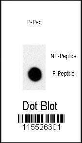
Dot blot analysis of anti-Phospho-MCAM-pY641 Antibody on nitrocellulose membrane. 50ng of Phospho-peptide or Non Phospho-peptide per dot were adsorbed. Antibody working concentrations are 0.5ug per ml.| Product Name | Phospho-MCAM-pY641 Antibody |
|---|---|
| Antibody Type | Primary Antibodies |
| Modification | p-Y641 |
| Clonality | Polyclonal |
|---|---|
| Isotype | Ig |
| Host Species | Rabbit |
| Tested Applications | DB |
| DB:1:500 |
|
| Species Reactivity | Human |
| Concentration | 1mg/ml |
| Gene Synonyms | MUC18 |
|---|---|
| Alternative Names | MCAM MUC18 Cell surface glycoprotein MUC18 Cell surface glycoprotein P1H12 Melanoma cell adhesion molecule Melanoma-associated antigen A32 Melanoma-associated antigen MUC18 S-endo 1 endothelial-associated antigen CD_antigen=CD146 Flags: Precursor |
| Molecular Weight(MW) | 71607 Da |
| Function | Plays a role in cell adhesion, and in cohesion of the endothelial monolayer at intercellular junctions in vascular tissue. Its expression may allow melanoma cells to interact with cellular elements of the vascular system, thereby enhancing hematogeneous tumor spread. Could be an adhesion molecule active in neural crest cells during embryonic development. Acts as surface receptor that triggers tyrosine phosphorylation of FYN and PTK2/FAK1, and a transient increase in the intracellular calcium concentration |
| Tissue Specificity | Detected in endothelial cells in vascular tissue throughout the body. May appear at the surface of neural crest cells during their embryonic migration. Appears to be limited to vascular smooth muscle in normal adult tissues Associated with tumor progression and the development of metastasis in human malignant melanoma. Expressed most strongly on metastatic lesions and advanced primary tumors and is only rarely detected in benign melanocytic nevi and thin primary melanomas with a low probability of metastasis |
| Cellular Localization | Membrane; Single-pass type I membrane protein |
Application
Dot blot analysis of anti-Phospho-MCAM-pY641 Antibody on nitrocellulose membrane. 50ng of Phospho-peptide or Non Phospho-peptide per dot were adsorbed. Antibody working concentrations are 0.5ug per ml.| Application Notes | DB:1:500 |
|---|
| Form | Liquid |
|---|---|
| Storage Instructions | For short-term storage, store at 4° C. For long-term storage, aliquot and store at -20ºC or below. Avoid multiple freeze-thaw cycles. |
| Storage Buffer | Purified polyclonal antibody supplied in PBS with 0.09% (W/V) sodium azide. This antibody is purified through a protein A column, followed by two-step phosphospecific peptide affinity purification. |


