Application
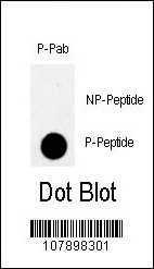
Dot blot analysis of anti-Phospho-MYT1-T495 Antibody on nitrocellulose membrane. 50ng of Phospho-peptide or Non Phospho-peptide per dot were adsorbed. Antibody working concentrations are 0.5ug per ml.Application
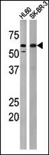
The anti-Phospho-MYT1-T495 Antibody is used in Western blot to detect Phospho-MYT1-T495 in HL60 (left) and SK-BR-3 (right) tissue lysates.Application
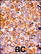
Formalin-fixed and paraffin-embedded human cancer tissue reacted with the primary antibody, which was peroxidase-conjugated to the secondary antibody, followed by AEC staining. This data demonstrates the use of this antibody for immunohistochemistry; clinical relevance has not been evaluated. BC = breast carcinoma; HC = hepatocarcinoma.| Product Name | Phospho-MYT1-T495 Antibody |
|---|---|
| Antibody Type | Primary Antibodies |
| Modification | p-T495 |
| Clonality | Polyclonal |
|---|---|
| Isotype | Ig |
| Host Species | Rabbit |
| Tested Applications | DBWBIHC |
| DB:1:500 WB |
|
| Species Reactivity | Human |
| Concentration | 1mg/ml |
| Gene Synonyms | MYT1 |
|---|---|
| Alternative Names | PKMYT1 MYT1 Membrane-associated tyrosine- and threonine-specific cdc2-inhibitory kinase Myt1 kinase |
| Molecular Weight(MW) | 54521 Da |
| Function | Acts as a negative regulator of entry into mitosis (G2 to M transition) by phosphorylation of the CDK1 kinase specifically when CDK1 is complexed to cyclins. Mediates phosphorylation of CDK1 predominantly on 'Thr-14'. Also involved in Golgi fragmentation. May be involved in phosphorylation of CDK1 on 'Tyr-15' to a lesser degree, however tyrosine kinase activity is unclear and may be indirect. May be a downstream target of Notch signaling pathway during eye development |
| Tissue Specificity | This Phospho-MYT1-T495 antibody is generated from rabbits immunized with a KLH conjugated synthetic phosphopeptide corresponding to amino acid residues surrounding T495 of human MYT1. |
| Cellular Localization | Endoplasmic reticulum membrane; Peripheral membrane protein. Golgi apparatus membrane; Peripheral membrane protein |
| Entrez Gene | 9088 |
|---|
Application
Dot blot analysis of anti-Phospho-MYT1-T495 Antibody on nitrocellulose membrane. 50ng of Phospho-peptide or Non Phospho-peptide per dot were adsorbed. Antibody working concentrations are 0.5ug per ml.
Application
The anti-Phospho-MYT1-T495 Antibody is used in Western blot to detect Phospho-MYT1-T495 in HL60 (left) and SK-BR-3 (right) tissue lysates.
Application
Formalin-fixed and paraffin-embedded human cancer tissue reacted with the primary antibody, which was peroxidase-conjugated to the secondary antibody, followed by AEC staining. This data demonstrates the use of this antibody for immunohistochemistry; clinical relevance has not been evaluated. BC = breast carcinoma; HC = hepatocarcinoma.| Application Notes | DB:1:500 WB |
|---|
| Form | Liquid |
|---|---|
| Storage Instructions | For short-term storage, store at 4° C. For long-term storage, aliquot and store at -20ºC or below. Avoid multiple freeze-thaw cycles. |
| Storage Buffer | Purified polyclonal antibody supplied in PBS with 0.09% (W/V) sodium azide. This antibody is first purified by protein G affinity chromatography. Then, the antibody fraction is peptide affinity purified in a 2-step procedure with control and phosphorylated peptides. The phospho-specific antibody is eluted with high and low pH buffers and neutralized immediately, followed by dialysis against PBS. |


