Application
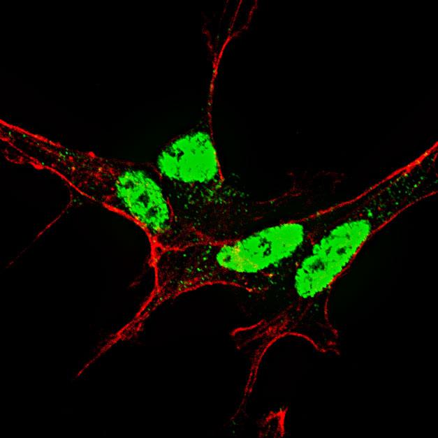
Fluorescent confocal image of SY5Y cells stained with AP3649a Phospho-Nanog-S71 antibody. SY5Y cells were fixed with 4% PFA (20 min), permeabilized with Triton X-100 (0.2%, 30 min), then incubated with AP3649a Phospho-Nanog-S71 primary antibody (1:500, 2 h at room temperature). For secondary antibody, Alexa Fluor® 488 conjugated donkey anti-rabbit antibody (green) was used (1:1000, 1h). Cytoplasmic actin was counterstained with Alexa Fluor® 555 (red) conjugated Phalloidin (5.25 μM, 25 min). Phospho-Nanog immunoreactivity is localized very specifically to the nuclei of the SY5Y cells.Application
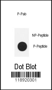
Dot blot analysis of anti-Phospho-Nanog-S71 Antibody on nitrocellulose membrane. 50ng of Phospho-peptide or Non Phospho-peptide per dot were adsorbed. Antibody working concentrations are 0.5ug per ml.| Product Name | Phospho-Nanog-S71 Antibody |
|---|---|
| Antibody Type | Primary Antibodies |
| Antigen Alias | NANOG; Homeobox protein NANOG; Homeobox transcription factor Nanog |
| Modification | p-S71 |
| Clonality | Polyclonal |
|---|---|
| Isotype | Ig |
| Host Species | Rabbit |
| Tested Applications | IFDB |
| IF:1:10~50 DB |
|
| Species Reactivity | Human |
| Concentration | 1mg/ml |
| Alternative Names | NANOG Homeobox protein NANOG Homeobox transcription factor Nanog |
|---|---|
| Molecular Weight(MW) | 34620 Da |
| Function | Transcription regulator involved in inner cell mass and embryonic stem (ES) cells proliferation and self-renewal. Imposes pluripotency on ES cells and prevents their differentiation towards extraembryonic endoderm and trophectoderm lineages. Blocks bone morphogenetic protein-induced mesoderm differentiation of ES cells by physically interacting with SMAD1 and interfering with the recruitment of coactivators to the active SMAD transcriptional complexes. Acts as a transcriptional activator or repressor. Binds optimally to the DNA consensus sequence 5'-TAAT[GT][GT]-3' or 5'- [CG][GA][CG]C[GC]ATTAN[GC]-3'. Able to autorepress its expression in differentiating (ES) cells: binds to its own promoter following interaction with ZNF281/ZFP281, leading to recruitment of the NuRD complex and subsequent repression of expression. When overexpressed, promotes cells to enter into S phase and proliferation |
| Tissue Specificity | Expressed in testicular carcinoma and derived germ cell tumors (at protein level). Expressed in fetal gonads, ovary and testis. Also expressed in ovary teratocarcinoma cell line and testicular embryonic carcinoma. Not expressed in many somatic organs and oocytes |
| Cellular Localization | Nucleus. |
| Entrez Gene | 79923 |
|---|
Application
Fluorescent confocal image of SY5Y cells stained with AP3649a Phospho-Nanog-S71 antibody. SY5Y cells were fixed with 4% PFA (20 min), permeabilized with Triton X-100 (0.2%, 30 min), then incubated with AP3649a Phospho-Nanog-S71 primary antibody (1:500, 2 h at room temperature). For secondary antibody, Alexa Fluor® 488 conjugated donkey anti-rabbit antibody (green) was used (1:1000, 1h). Cytoplasmic actin was counterstained with Alexa Fluor® 555 (red) conjugated Phalloidin (5.25 μM, 25 min). Phospho-Nanog immunoreactivity is localized very specifically to the nuclei of the SY5Y cells.
Application
Dot blot analysis of anti-Phospho-Nanog-S71 Antibody on nitrocellulose membrane. 50ng of Phospho-peptide or Non Phospho-peptide per dot were adsorbed. Antibody working concentrations are 0.5ug per ml.| Application Notes | IF:1:10~50 DB |
|---|
| Form | Liquid |
|---|---|
| Storage Instructions | For short-term storage, store at 4° C. For long-term storage, aliquot and store at -20ºC or below. Avoid multiple freeze-thaw cycles. |
| Storage Buffer | Purified polyclonal antibody supplied in PBS with 0.09% (W/V) sodium azide. This antibody is purified through a protein A column, followed by two-step phosphospecific peptide affinity purification. |


