Application
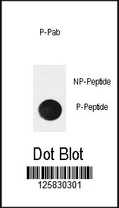
Dot blot analysis of anti-Phospho-NFKBIA (Ser32) Antibody Phospho-specific Pab on nitrocellulose membrane. 50ng of Phospho-peptide or Non Phospho-peptide per dot were adsorbed. Antibody working concentrations are 0.5ug per ml.| Product Name | Phospho-NFKBIA (Ser32) Antibody |
|---|---|
| Antibody Type | Primary Antibodies |
| Antigen Alias | NFKBIA; IKBA; MAD3; NFKBI; NF-kappa-B inhibitor alpha; I-kappa-B-alpha; Major histocompatibility complex enhancer-binding protein MAD3 |
| Modification | p-Ser32 |
| Clonality | Polyclonal |
|---|---|
| Isotype | Ig |
| Host Species | Rabbit |
| Tested Applications | DB |
| DB:1:500 |
|
| Species Reactivity | Human |
| Concentration | 1mg/ml |
| Gene Synonyms | IKBA MAD3 NFKBI |
|---|---|
| Alternative Names | NFKBIA IKBA MAD3 NFKBI NF-kappa-B inhibitor alpha I-kappa-B-alpha Major histocompatibility complex enhancer-binding protein MAD3 |
| Molecular Weight(MW) | 35609 Da |
| Function | Inhibits the activity of dimeric NF-kappa-B/REL complexes by trapping REL dimers in the cytoplasm through masking of their nuclear localization signals. On cellular stimulation by immune and proinflammatory responses, becomes phosphorylated promoting ubiquitination and degradation, enabling the dimeric RELA to translocate to the nucleus and activate transcription |
| Tissue Specificity | This Phospho-NFKBIA (Ser32) antibody is generated from rabbits immunized with a KLH conjugated synthetic phosphopeptide between amino acids 20-50 surrounding S32 of human NFKBIA. |
| Cellular Localization | Cytoplasm. Nucleus. Note=Shuttles between the nucleus and the cytoplasm by a nuclear localization signal (NLS) and a CRM1-dependent nuclear export (By similarity) |
| Entrez Gene | 4792 |
|---|
Application
Dot blot analysis of anti-Phospho-NFKBIA (Ser32) Antibody Phospho-specific Pab on nitrocellulose membrane. 50ng of Phospho-peptide or Non Phospho-peptide per dot were adsorbed. Antibody working concentrations are 0.5ug per ml.| Application Notes | DB:1:500 |
|---|
| Form | Liquid |
|---|---|
| Storage Instructions | For short-term storage, store at 4° C. For long-term storage, aliquot and store at -20ºC or below. Avoid multiple freeze-thaw cycles. |
| Storage Buffer | Purified polyclonal antibody supplied in PBS with 0.09% (W/V) sodium azide. This antibody is purified through a protein A column, followed by two-step phosphospecific peptide affinity purification. |


