Application
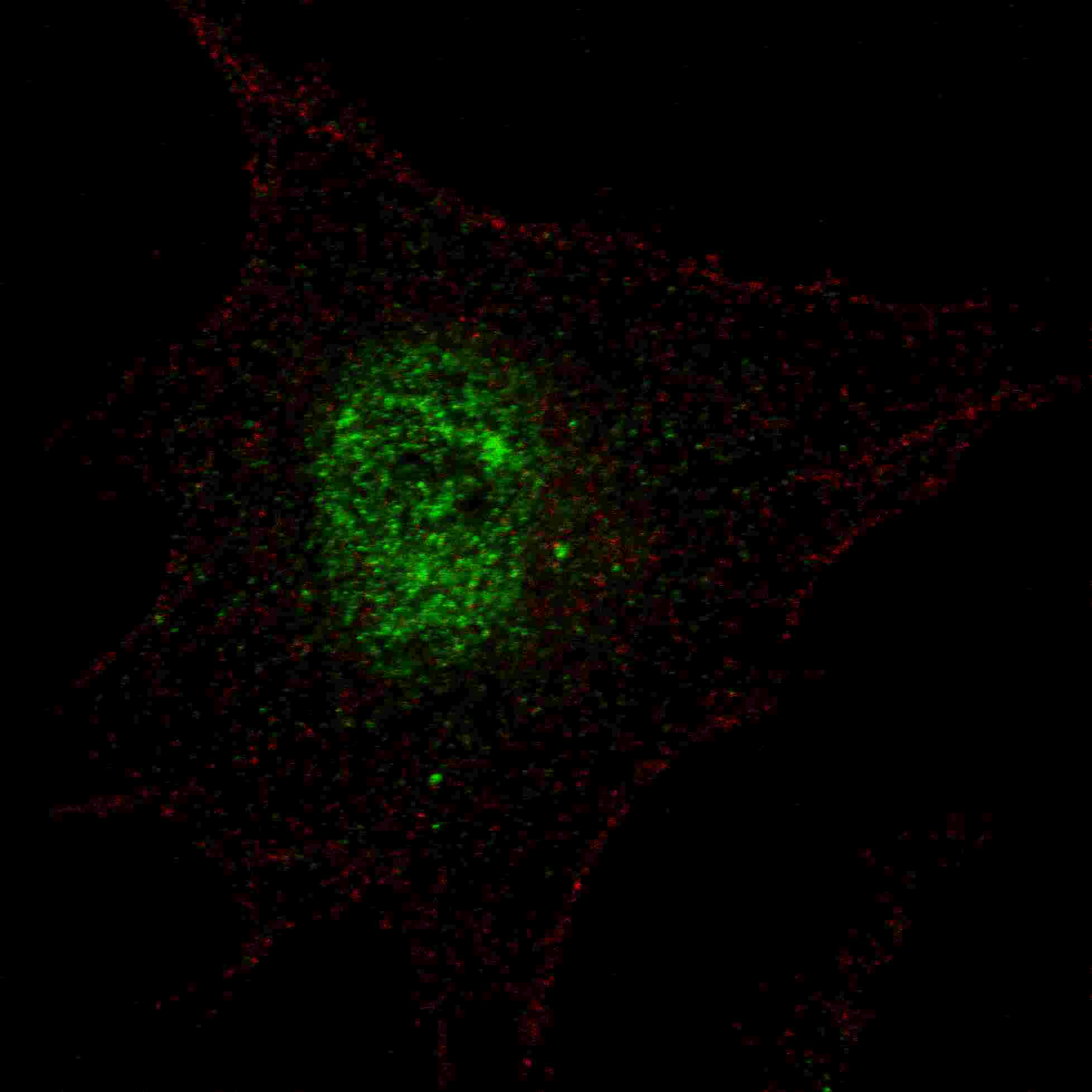
Fluorescent confocal image of SY5Y cells stained with phospho-PDX1-T11 antibody. SY5Y cells were fixed with 4% PFA (20 min), permeabilized with Triton X-100 (0.2%, 30 min). Cells were then incubated with AP3669a phospho-PDX1-T11 primary antibody (1:100, 2 h at room temperature). For secondary antibody, Alexa Fluor® 488 conjugated donkey anti-rabbit antibody (green) was used (1:1000, 1h). Nuclei were counterstained with Hoechst 33342 (blue) (10 μg/ml, 5 min). Note the highly specific localization of the phospho-PDX1 immunosignal mainly to the nucleus.Application
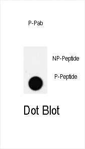
Dot blot analysis of PDX1 Antibody (T11) Pab on nitrocellulose membrane. 50ng of Phospho-peptide or Non Phospho-peptide per dot were adsorbed. Antibody working concentrations are 0.5ug per ml.| Product Name | Phospho-PDX1-T11 Antibody |
|---|---|
| Antibody Type | Primary Antibodies |
| Antigen Alias | PDX1; IPF1; Pancreas/duodenum homeobox protein 1; Glucose-sensitive factor; Insulin promoter factor 1; Insulin upstream factor 1; Islet/duodenum homeobox-1; Somatostatin-transactivating factor 1 |
| Modification | p-T11 |
| Clonality | Polyclonal |
|---|---|
| Isotype | Ig |
| Host Species | Rabbit |
| Tested Applications | IFDB |
| IF:1:100 DB |
|
| Species Reactivity | Human |
| Concentration | 1mg/ml |
| Gene Synonyms | IPF1 STF1 |
|---|---|
| Alternative Names | PDX1 IPF1 Pancreas/duodenum homeobox protein 1 Glucose-sensitive factor Insulin promoter factor 1 Insulin upstream factor 1 Islet/duodenum homeobox-1 Somatostatin-transactivating factor 1 |
| Molecular Weight(MW) | 30771 Da |
| Function | Activates insulin, somatostatin, glucokinase, islet amyloid polypeptide and glucose transporter type 2 gene transcription. Particularly involved in glucose-dependent regulation of insulin gene transcription. As part of a PDX1:PBX1b:MEIS2b complex in pancreatic acinar cells is involved in the transcriptional activation of the ELA1 enhancer; the complex binds to the enhancer B element and cooperates with the transcription factor 1 complex (PTF1) bound to the enhancer A element. Binds preferentially the DNA motif 5'-[CT]TAAT[TG]-3' During development, specifies the early pancreatic epithelium, permitting its proliferation, branching and subsequent differentiation. At adult stage, required for maintaining the hormone-producing phenotype of the beta-cell |
| Tissue Specificity | Duodenum and pancreas (Langerhans islet beta cells and small subsets of endocrine non-beta-cells, at low levels in acinar cells) |
| Cellular Localization | Nucleus. Cytoplasm, cytosol (By similarity). |
Application
Fluorescent confocal image of SY5Y cells stained with phospho-PDX1-T11 antibody. SY5Y cells were fixed with 4% PFA (20 min), permeabilized with Triton X-100 (0.2%, 30 min). Cells were then incubated with AP3669a phospho-PDX1-T11 primary antibody (1:100, 2 h at room temperature). For secondary antibody, Alexa Fluor® 488 conjugated donkey anti-rabbit antibody (green) was used (1:1000, 1h). Nuclei were counterstained with Hoechst 33342 (blue) (10 μg/ml, 5 min). Note the highly specific localization of the phospho-PDX1 immunosignal mainly to the nucleus.
Application
Dot blot analysis of PDX1 Antibody (T11) Pab on nitrocellulose membrane. 50ng of Phospho-peptide or Non Phospho-peptide per dot were adsorbed. Antibody working concentrations are 0.5ug per ml.| Application Notes | IF:1:100 DB |
|---|
| Form | Liquid |
|---|---|
| Storage Instructions | For short-term storage, store at 4° C. For long-term storage, aliquot and store at -20ºC or below. Avoid multiple freeze-thaw cycles. |
| Storage Buffer | Purified polyclonal antibody supplied in PBS with 0.09% (W/V) sodium azide. This antibody is purified through a protein A column, followed by two-step phosphospecific peptide affinity purification. |


