Application
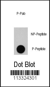
Dot blot analysis of anti-PIK3R1-pY556 Phospho-specific Pab (RB13324) on nitrocellulose membrane. 50ng of Phospho-peptide or Non Phospho-peptide per dot were adsorbed. Antibody working concentrations are 0.5ug per ml.| Product Name | Phospho-PIK3R1-Y556 Antibody |
|---|---|
| Antibody Type | Primary Antibodies |
| Antigen Alias | PIK3R1; GRB1; Phosphatidylinositol 3-kinase regulatory subunit alpha; Phosphatidylinositol 3-kinase 85 kDa regulatory subunit alpha |
| Modification | p-Y556 |
| Clonality | Polyclonal |
|---|---|
| Isotype | Ig |
| Host Species | Rabbit |
| Tested Applications | DB |
| DB:1:500 |
|
| Species Reactivity | Human |
| Concentration | 1mg/ml |
| Gene Synonyms | GRB1 |
|---|---|
| Alternative Names | PIK3R1 GRB1 Phosphatidylinositol 3-kinase regulatory subunit alpha Phosphatidylinositol 3-kinase 85 kDa regulatory subunit alpha |
| Molecular Weight(MW) | 83598 Da |
| Function | Binds to activated (phosphorylated) protein-Tyr kinases, through its SH2 domain, and acts as an adapter, mediating the association of the p110 catalytic unit to the plasma membrane Necessary for the insulin-stimulated increase in glucose uptake and glycogen synthesis in insulin-sensitive tissues. Plays an important role in signaling in response to FGFR1, FGFR2, FGFR3, FGFR4, KITLG/SCF, KIT, PDGFRA and PDGFRB. Likewise, plays a role in ITGB2 signaling |
| Tissue Specificity | Isoform 2 is expressed in skeletal muscle and brain, and at lower levels in kidney and cardiac muscle. Isoform 2 and isoform 4 are present in skeletal muscle (at protein level) |
| Entrez Gene | 5295 |
|---|
Application
Dot blot analysis of anti-PIK3R1-pY556 Phospho-specific Pab (RB13324) on nitrocellulose membrane. 50ng of Phospho-peptide or Non Phospho-peptide per dot were adsorbed. Antibody working concentrations are 0.5ug per ml.| Application Notes | DB:1:500 |
|---|
| Form | Liquid |
|---|---|
| Storage Instructions | For short-term storage, store at 4° C. For long-term storage, aliquot and store at -20ºC or below. Avoid multiple freeze-thaw cycles. |
| Storage Buffer | Purified rabbit polyclonal antibody supplied in PBS with 0.09% (W/V) sodium azide. Then, the antibody fraction is peptide affinity purified in a 2-step procedure with the control and phosphorylated peptides. The phospho-specific antibody is eluted with high and low pH buffers and neutralized immediately, followed by dialysis against PBS. |


