Application
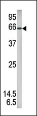
Western blot analysis of anti-Phospho-STAT1 Pab in mouse brain tissue lysate (35ug/lane). Phospho-STAT1(arrow) was detected using the purified Pab (1:80 dilution).Application
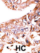
Formalin-fixed and paraffin-embedded human cancer tissue reacted with the primary antibody, which was peroxidase-conjugated to the secondary antibody, followed by AEC staining. This data demonstrates the use of this antibody for immunohistochemistry; clinical relevance has not been evaluated. BC = breast carcinoma; HC = hepatocarcinoma.| Product Name | Phospho-STAT1-S727 Antibody |
|---|---|
| Antibody Type | Primary Antibodies |
| Antigen Alias | STAT1; Signal transducer and activator of transcription 1-alpha/beta; Transcription factor ISGF-3 components p91/p84 |
| Modification | p-S727 |
| Clonality | Polyclonal |
|---|---|
| Isotype | Ig |
| Host Species | Rabbit |
| Tested Applications | WBIHC |
| WB:1:100~500 IHC |
|
| Species Reactivity | Human |
| Concentration | 1mg/ml |
| Alternative Names | STAT1 Signal transducer and activator of transcription 1-alpha/beta Transcription factor ISGF-3 components p91/p84 |
|---|---|
| Molecular Weight(MW) | 87335 Da |
| Function | Signal transducer and transcription activator that mediates cellular responses to interferons (IFNs), cytokine KITLG/SCF and other cytokines and growth factors. Following type I IFN (IFN-alpha and IFN-beta) binding to cell surface receptors, signaling via protein kinases leads to activation of Jak kinases (TYK2 and JAK1) and to tyrosine phosphorylation of STAT1 and STAT2. The phosphorylated STATs dimerize, associate with ISGF3G/IRF-9 to form a complex termed ISGF3 transcription factor, that enters the nucleus. ISGF3 binds to the IFN stimulated response element (ISRE) to activate the transcription of interferon stimulated genes, which drive the cell in an antiviral state. In response to type II IFN (IFN-gamma), STAT1 is tyrosine- and serine-phosphorylated. It then forms a homodimer termed IFN- gamma-activated factor (GAF), migrates into the nucleus and binds to the IFN gamma activated sequence (GAS) to drive the expression of the target genes, inducing a cellular antiviral state. Becomes activated in response to KITLG/SCF and KIT signaling. May mediate cellular responses to activated FGFR1, FGFR2, FGFR3 and FGFR4 |
| Tissue Specificity | This Phospho-STAT1-S727 antibody is generated from rabbits immunized with a KLH conjugated synthetic phosphopeptide corresponding to amino acid residues surrounding S727 of human STAT1. |
| Cellular Localization | Cytoplasm. Nucleus. Note=Translocated into the nucleus upon tyrosine phosphorylation and dimerization, in response to IFN-gamma and signaling by activated FGFR1, FGFR2, FGFR3 or FGFR4 |
| Entrez Gene | 6772 |
|---|
Application
Western blot analysis of anti-Phospho-STAT1 Pab in mouse brain tissue lysate (35ug/lane). Phospho-STAT1(arrow) was detected using the purified Pab (1:80 dilution).
Application
Formalin-fixed and paraffin-embedded human cancer tissue reacted with the primary antibody, which was peroxidase-conjugated to the secondary antibody, followed by AEC staining. This data demonstrates the use of this antibody for immunohistochemistry; clinical relevance has not been evaluated. BC = breast carcinoma; HC = hepatocarcinoma.| Application Notes | WB:1:100~500 IHC |
|---|
| Form | Liquid |
|---|---|
| Storage Instructions | For short-term storage, store at 4° C. For long-term storage, aliquot and store at -20ºC or below. Avoid multiple freeze-thaw cycles. |
| Storage Buffer | Purified polyclonal antibody supplied in PBS with 0.09% (W/V) sodium azide. This antibody is first purified by protein G affinity chromatography. Then, the antibody fraction is peptide affinity purified in a 2-step procedure with control and phosphorylated peptides. The phospho-specific antibody is eluted with high and low pH buffers and neutralized immediately, followed by dialysis against PBS. |


