Application
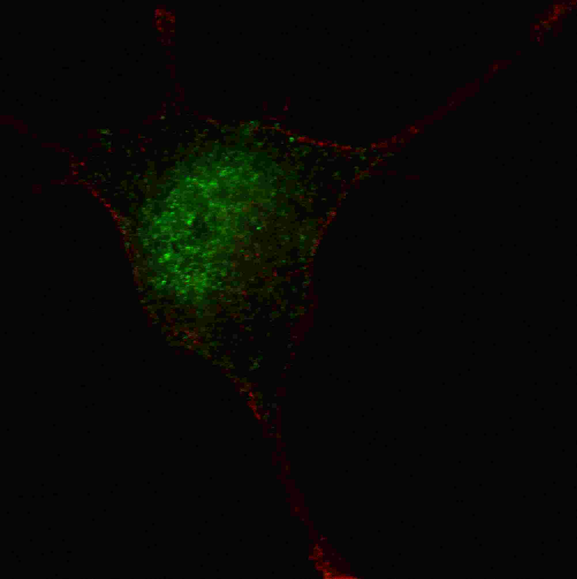
Fluorescent confocal image of SY5Y cells stained with phospho-STAT3-S727 antibody. SY5Y cells were fixed with 4% PFA (20 min), permeabilized with Triton X-100 (0.2%, 30 min). Cells were then incubated with AP1534a phospho-STAT3-S727 primary antibody (1:100, 2 h at room temperature). For secondary antibody, Alexa Fluor® 488 conjugated donkey anti-rabbit antibody (green) was used (1:1000, 1h). Nuclei were counterstained with Hoechst 33342 (blue) (10 μg/ml, 5 min). Note the highly specific localization of the phospho-STAT3-S727 immunosignal to the nucleus and cytoplasm, supported by Human Protein Atlas Data (http://www.proteinatlas.org/ENSG00000168610).Application
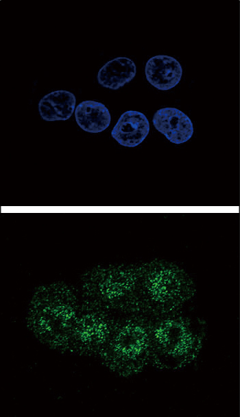
Confocal immunofluorescent analysis of Phospho-STAT3-S727 Antibody(Cat#AP1534a) with HepG2 cell followed by Alexa Fluor 488-conjugated goat anti-rabbit lgG (green). DAPI was used to stain the cell nuclear (blue).| Product Name | Phospho-STAT3-S727 Antibody |
|---|---|
| Antibody Type | Primary Antibodies |
| Antigen Alias | STAT3; APRF; Signal transducer and activator of transcription 3; Acute-phase response factor |
| Modification | p-S727 |
| Clonality | Polyclonal |
|---|---|
| Isotype | Ig |
| Host Species | Rabbit |
| Tested Applications | IF |
| IF:1:10~50 |
|
| Species Reactivity | Human |
| Concentration | 1mg/ml |
| Gene Synonyms | APRF |
|---|---|
| Alternative Names | STAT3 APRF Signal transducer and activator of transcription 3 Acute-phase response factor |
| Molecular Weight(MW) | 88068 Da |
| Function | Signal transducer and transcription activator that mediates cellular responses to interleukins, KITLG/SCF and other growth factors. May mediate cellular responses to activated FGFR1, FGFR2, FGFR3 and FGFR4. Binds to the interleukin-6 (IL-6)- responsive elements identified in the promoters of various acute- phase protein genes. Activated by IL31 through IL31RA |
| Tissue Specificity | Heart, brain, placenta, lung, liver, skeletal muscle, kidney and pancreas |
| Cellular Localization | Cytoplasm. Nucleus. Note=Shuttles between the nucleus and the cytoplasm. Translocated into the nucleus upon tyrosine phosphorylation and dimerization, in response to signaling by activated FGFR1, FGFR2, FGFR3 or FGFR4. Constitutive nuclear presence is independent of tyrosine phosphorylation Predominantly present in the cytoplasm without stimuli. Upon leukemia inhibitory factor (LIF) stimulation, accumulates in the nucleus. The complex composed of BART and ARL2 plays an important role in the nuclear translocation and retention of STAT3 Identified in a complex with LYN and PAG1 |
| Entrez Gene | 6774 |
|---|
Application
Fluorescent confocal image of SY5Y cells stained with phospho-STAT3-S727 antibody. SY5Y cells were fixed with 4% PFA (20 min), permeabilized with Triton X-100 (0.2%, 30 min). Cells were then incubated with AP1534a phospho-STAT3-S727 primary antibody (1:100, 2 h at room temperature). For secondary antibody, Alexa Fluor® 488 conjugated donkey anti-rabbit antibody (green) was used (1:1000, 1h). Nuclei were counterstained with Hoechst 33342 (blue) (10 μg/ml, 5 min). Note the highly specific localization of the phospho-STAT3-S727 immunosignal to the nucleus and cytoplasm, supported by Human Protein Atlas Data (http://www.proteinatlas.org/ENSG00000168610).
Application
Confocal immunofluorescent analysis of Phospho-STAT3-S727 Antibody(Cat#AP1534a) with HepG2 cell followed by Alexa Fluor 488-conjugated goat anti-rabbit lgG (green). DAPI was used to stain the cell nuclear (blue).| Application Notes | IF:1:10~50 |
|---|
| Form | Liquid |
|---|---|
| Storage Instructions | For short-term storage, store at 4° C. For long-term storage, aliquot and store at -20ºC or below. Avoid multiple freeze-thaw cycles. |
| Storage Buffer | Purified polyclonal antibody supplied in PBS with 0.09% (W/V) sodium azide. This antibody is first purified by protein G affinity chromatography. Then, the antibody fraction is peptide affinity purified in a 2-step procedure with control and phosphorylated peptides. The phospho-specific antibody is eluted with high and low pH buffers and neutralized immediately, followed by dialysis against PBS. |


