Application
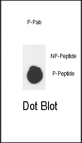
Dot blot analysis of anti-Phospho-Wee1-S53 Antibody on nitrocellulose membrane. 50ng of Phospho-peptide or Non Phospho-peptide per dot were adsorbed. Antobodies working concentration was 0.5ug per ml.| Product Name | Phospho-Wee1-S53 Antibody |
|---|---|
| Antibody Type | Primary Antibodies |
| Antigen Alias | WEE1; Wee1-like protein kinase; Wee1A kinase |
| Modification | p-S53 |
| Clonality | Polyclonal |
|---|---|
| Isotype | Ig |
| Host Species | Rabbit |
| Tested Applications | DB |
| DB:0.388888888889 |
|
| Species Reactivity | Human |
| Concentration | 1mg/ml |
| Alternative Names | WEE1 Wee1-like protein kinase Wee1A kinase |
|---|---|
| Molecular Weight(MW) | 71597 Da |
| Function | Acts as a negative regulator of entry into mitosis (G2 to M transition) by protecting the nucleus from cytoplasmically activated cyclin B1-complexed CDK1 before the onset of mitosis by mediating phosphorylation of CDK1 on 'Tyr-15'. Specifically phosphorylates and inactivates cyclin B1-complexed CDK1 reaching a maximum during G2 phase and a minimum as cells enter M phase Phosphorylation of cyclin B1-CDK1 occurs exclusively on 'Tyr-15' and phosphorylation of monomeric CDK1 does not occur. Its activity increases during S and G2 phases and decreases at M phase when it is hyperphosphorylated. A correlated decrease in protein level occurs at M/G1 phase, probably due to its degradation |
| Tissue Specificity | This Phospho-Wee1-S53 antibody is generated from rabbits immunized with a KLH conjugated synthetic phosphopeptide corresponding to amino acid residues surrounding S53 of human WEE1 |
| Cellular Localization | Nucleus. |
| Entrez Gene | 7465 |
|---|
Application
Dot blot analysis of anti-Phospho-Wee1-S53 Antibody on nitrocellulose membrane. 50ng of Phospho-peptide or Non Phospho-peptide per dot were adsorbed. Antobodies working concentration was 0.5ug per ml.| Application Notes | DB:0.388888888889 |
|---|
| Form | Liquid |
|---|---|
| Storage Instructions | For short-term storage, store at 4° C. For long-term storage, aliquot and store at -20ºC or below. Avoid multiple freeze-thaw cycles. |
| Storage Buffer | Purified polyclonal antibody supplied in PBS with 0.09% (W/V) sodium azide. This antibody is first purified by protein G affinity chromatography. Then, the antibody fraction is peptide affinity purified in a 2-step procedure with control and phosphorylated peptides. The phospho-specific antibody is eluted with high and low pH buffers and neutralized immediately, followed by dialysis against PBS. |


