Application
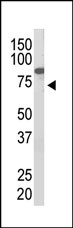
Western blot analysis of PIASx1 polyclonal antibody with bacterial and HL-60 cell lysates. Lane A: endogenous PIASx1 in HL-60 cells; Lane B: bacteria expressing recombinant GST-PIASx1 fusion protein. PIASx1 was detected using purified Pab. Secondary HRP-anti-rabbit was used for signal visualization with chemiluminescence.Application
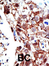
Formalin-fixed and paraffin-embedded human cancer tissue reacted with the primary antibody, which was peroxidase-conjugated to the secondary antibody, followed by AEC staining. This data demonstrates the use of this antibody for immunohistochemistry; clinical relevance has not been evaluated. BC = breast carcinoma; HC = hepatocarcinoma.| Product Name | PIAS2 (PIASx1) Antibody (C-term) |
|---|---|
| Antibody Type | Primary Antibodies |
| Antigen Alias | PIAS2; PIASX; E3 SUMO-protein ligase PIAS2; Androgen receptor-interacting protein 3; DAB2-interacting protein; Msx-interacting zinc finger protein; PIAS-NY protein; Protein inhibitor of activated STAT x; Protein inhibitor of activated STAT2 |
| Clonality | Polyclonal |
|---|---|
| Isotype | Ig |
| Host Species | Rabbit |
| Tested Applications | WBIHC |
| WB:1:100~500 IHC |
|
| Species Reactivity | Human |
| Concentration | 1mg/ml |
| Gene Synonyms | PIASX |
|---|---|
| Alternative Names | PIAS2 PIASX E3 SUMO-protein ligase PIAS2 Androgen receptor-interacting protein 3 DAB2-interacting protein Msx-interacting zinc finger protein PIAS-NY protein Protein inhibitor of activated STAT x Protein inhibitor of activated STAT2 |
| Molecular Weight(MW) | 68240 Da |
| Function | Functions as an E3-type small ubiquitin-like modifier (SUMO) ligase, stabilizing the interaction between UBE2I and the substrate, and as a SUMO-tethering factor. Plays a crucial role as a transcriptional coregulator in various cellular pathways, including the STAT pathway, the p53 pathway and the steroid hormone signaling pathway. The effects of this transcriptional coregulation, transactivation or silencing may vary depending upon the biological context and the PIAS2 isoform studied. However, it seems to be mostly involved in gene silencing. Binds to sumoylated ELK1 and enhances its transcriptional activity by preventing recruitment of HDAC2 by ELK1, thus reversing SUMO-mediated repression of ELK1 transactivation activity. Isoform PIAS2-beta, but not isoform PIAS2-alpha, promotes MDM2 sumoylation. Isoform PIAS2-alpha promotes PARK7 sumoylation. Isoform PIAS2-beta promotes NCOA2 sumoylation more efficiently than isoform PIAS2- alpha |
| Tissue Specificity | Mainly expressed in testis. Isoform 3 is expressed predominantly in adult testis, weakly in pancreas, embryonic testis and sperm, and at very low levels in other organs |
| Cellular Localization | Nucleus speckle. Nucleus, PML body. Note=Colocalizes at least partially with promyelocytic leukemia nuclear bodies (PML NBs) |
| Entrez Gene | 9063 |
|---|
Application
Western blot analysis of PIASx1 polyclonal antibody with bacterial and HL-60 cell lysates. Lane A: endogenous PIASx1 in HL-60 cells; Lane B: bacteria expressing recombinant GST-PIASx1 fusion protein. PIASx1 was detected using purified Pab. Secondary HRP-anti-rabbit was used for signal visualization with chemiluminescence.
Application
Formalin-fixed and paraffin-embedded human cancer tissue reacted with the primary antibody, which was peroxidase-conjugated to the secondary antibody, followed by AEC staining. This data demonstrates the use of this antibody for immunohistochemistry; clinical relevance has not been evaluated. BC = breast carcinoma; HC = hepatocarcinoma.| Application Notes | WB:1:100~500 IHC |
|---|
| Form | Liquid |
|---|---|
| Storage Instructions | For short-term storage, store at 4° C. For long-term storage, aliquot and store at -20ºC or below. Avoid multiple freeze-thaw cycles. |
| Storage Buffer | Purified polyclonal antibody supplied in PBS with 0.09% (W/V) sodium azide. This antibody is purified through a protein G column, eluted with high and low pH buffers and neutralized immediately, followed by dialysis against PBS. |


