Application
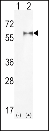
Western blot analysis of PIP5K1A (arrow) using rabbit polyclonal hPIP5K1A-R34 . 293 cell lysates (2 ug/lane) either nontransfected (Lane 1) or transiently transfected (Lane 2) with the PIP5K1A gene.| Product Name | PIP5K1A Antibody (N-term) |
|---|---|
| Antibody Type | Primary Antibodies |
| Antigen Alias | PIP5K1A; Phosphatidylinositol 4-phosphate 5-kinase type-1 alpha; 68 kDa type I phosphatidylinositol 4-phosphate 5-kinase alpha; Phosphatidylinositol 4-phosphate 5-kinase type I alpha |
| Clonality | Polyclonal |
|---|---|
| Isotype | Ig |
| Host Species | Rabbit |
| Tested Applications | WB |
| WB:1:100~500 |
|
| Species Reactivity | Human |
| Concentration | 1mg/ml |
| Alternative Names | PIP5K1A Phosphatidylinositol 4-phosphate 5-kinase type-1 alpha 68 kDa type I phosphatidylinositol 4-phosphate 5-kinase alpha Phosphatidylinositol 4-phosphate 5-kinase type I alpha |
|---|---|
| Molecular Weight(MW) | 62633 Da |
| Function | Catalyzes the phosphorylation of phosphatidylinositol 4- phosphate (PtdIns4P) to form phosphatidylinositol 4,5-bisphosphate (PtdIns(4,5)P2). PtdIns(4,5)P2 is involved in a variety of cellular processes and is the substrate to form phosphatidylinositol 3,4,5-trisphosphate (PtdIns(3,4,5)P3), another second messenger. The majority of PtdIns(4,5)P2 is thought to occur via type I phosphatidylinositol 4-phosphate 5-kinases given the abundance of PtdIns4P. Participates in a variety of cellular processes such as actin cytoskeleton organization, cell adhesion, migration and phagocytosis. Required for membrane ruffling formation, actin organization and focal adhesion formation during directional cell migration by controlling integrin-induced translocation of RAC1 to the plasma membrane Together with PIP5K1C is required for phagocytosis, but they regulate different types of actin remodeling at sequential steps Promotes particle ingestion by activating WAS that induces Arp2/3 dependent actin polymerization at the nascent phagocytic cup Together with PIP5K1B is required after stimulation of G-protein coupled receptors for stable platelet adhesion. Plays a role during calcium-induced keratinocyte differentiation. Recruited to the plasma membrane by the E-cadherin/beta-catenin complex where it provides the substrate PtdIns(4,5)P2 for the production of PtdIns(3,4,5)P3, diacylglycerol and inositol 1,4,5-trisphosphate that mobilize internal calcium and drive keratinocyte differentiation. Together with PIP5K1C have a role during embryogenesis. Functions also in the nucleus where acts as an activator of TUT1 adenylyltransferase activity in nuclear speckles, thereby regulating mRNA polyadenylation of a select set of mRNAs |
| Tissue Specificity | Highly expressed in heart, placenta, skeletal muscle, kidney and pancreas. Detected at lower levels in brain, lung and liver |
| Cellular Localization | Cell membrane. Cytoplasm (By similarity). Nucleus speckle. Cell projection, ruffle. Note=Colocalizes with RAC1 at actin-rich membrane ruffles. Localizes to nuclear speckles and associates with TUT1 to regulate polyadenylation of selected mRNAs |
| Entrez Gene | 8394 |
|---|
Application
Western blot analysis of PIP5K1A (arrow) using rabbit polyclonal hPIP5K1A-R34 . 293 cell lysates (2 ug/lane) either nontransfected (Lane 1) or transiently transfected (Lane 2) with the PIP5K1A gene.| Application Notes | WB:1:100~500 |
|---|
| Form | Liquid |
|---|---|
| Storage Instructions | For short-term storage, store at 4° C. For long-term storage, aliquot and store at -20ºC or below. Avoid multiple freeze-thaw cycles. |
| Storage Buffer | Purified polyclonal antibody supplied in PBS with 0.09% (W/V) sodium azide. This antibody is purified through a protein G column, eluted with high and low pH buffers and neutralized immediately, followed by dialysis against PBS. |


