Application
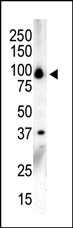
Western blot analysis of anti-PKCnu C-term Pab in NCI-H460 cell lysate. PKCnu (arrow) was detected using purified Pab. Secondary HRP-anti-rabbit was used for signal visualization with chemiluminescence.Application
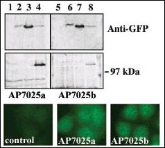
Upper panel, western blot analysis of GFP fusion protein expression in Panc-1 cells by using an anti-GFP antibody. Lanes 1 and 5: non-transfected cells; lanes 2 and 6: GFP-PKD-transfected cells; lanes 3 and 7: GFP-PKD2-transfected cells; lanes 4 and 8: GFP-PKD3 transfected cells. Center panel, western blot analysis of GFP fusion protein expression in Panc-1 cells by using PKD3 N-term (AP7025a) and C-term (AP7025b) antibodies. Lower panel, indirect immunofluorescence analysis of GFP-PKD3 fusion protein expression in Panc-1 cells by using AP7025a and AP7025b antibodies. Data courtesy of Dr. Osvaldo Rey, University of California Los Angeles.Application
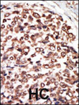
Formalin-fixed and paraffin-embedded human cancer tissue reacted with the primary antibody, which was peroxidase-conjugated to the secondary antibody, followed by AEC staining. This data demonstrates the use of this antibody for immunohistochemistry; clinical relevance has not been evaluated. BC = breast carcinoma; HC = hepatocarcinoma.| Product Name | PKC nu Antibody (C-term) |
|---|---|
| Antibody Type | Primary Antibodies |
| Antigen Alias | PRKD3; EPK2; PRKCN; Serine/threonine-protein kinase D3; Protein kinase C nu type; Protein kinase EPK2; nPKC-nu |
| Clonality | Polyclonal |
|---|---|
| Isotype | Ig |
| Host Species | Rabbit |
| Tested Applications | WBIHC |
| WB:1:100~500 IHC |
|
| Species Reactivity | Human |
| Concentration | 1mg/ml |
| Gene Synonyms | EPK2 PRKCN |
|---|---|
| Alternative Names | PRKD3 EPK2 PRKCN Serine/threonine-protein kinase D3 Protein kinase C nu type Protein kinase EPK2 nPKC-nu |
| Molecular Weight(MW) | 100471 Da |
| Function | Converts transient diacylglycerol (DAG) signals into prolonged physiological effects, downstream of PKC. Involved in resistance to oxidative stress (By similarity) |
| Tissue Specificity | Ubiquitous. |
| Cellular Localization | Cytoplasm. Membrane. Note=Translocation to the cell membrane is required for kinase activation |
| Entrez Gene | 23683 |
|---|
Application
Western blot analysis of anti-PKCnu C-term Pab in NCI-H460 cell lysate. PKCnu (arrow) was detected using purified Pab. Secondary HRP-anti-rabbit was used for signal visualization with chemiluminescence.
Application
Upper panel, western blot analysis of GFP fusion protein expression in Panc-1 cells by using an anti-GFP antibody. Lanes 1 and 5: non-transfected cells; lanes 2 and 6: GFP-PKD-transfected cells; lanes 3 and 7: GFP-PKD2-transfected cells; lanes 4 and 8: GFP-PKD3 transfected cells. Center panel, western blot analysis of GFP fusion protein expression in Panc-1 cells by using PKD3 N-term (AP7025a) and C-term (AP7025b) antibodies. Lower panel, indirect immunofluorescence analysis of GFP-PKD3 fusion protein expression in Panc-1 cells by using AP7025a and AP7025b antibodies. Data courtesy of Dr. Osvaldo Rey, University of California Los Angeles.
Application
Formalin-fixed and paraffin-embedded human cancer tissue reacted with the primary antibody, which was peroxidase-conjugated to the secondary antibody, followed by AEC staining. This data demonstrates the use of this antibody for immunohistochemistry; clinical relevance has not been evaluated. BC = breast carcinoma; HC = hepatocarcinoma.| Application Notes | WB:1:100~500 IHC |
|---|
| Form | Liquid |
|---|---|
| Storage Instructions | For short-term storage, store at 4° C. For long-term storage, aliquot and store at -20ºC or below. Avoid multiple freeze-thaw cycles. |
| Storage Buffer | Purified polyclonal antibody supplied in PBS with 0.09% (W/V) sodium azide. This antibody is purified through a protein G column, eluted with high and low pH buffers and neutralized immediately, followed by dialysis against PBS. |


