Application
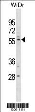
PLXDC1 Antibody (C-term) western blot analysis in WiDr cell line lysates (35ug/lane).This demonstrates the PLXDC1 antibody detected the PLXDC1 protein (arrow).| Product Name | PLXDC1 (C-term) |
|---|---|
| Antibody Type | Primary Antibodies |
| Antigen Alias | PLXDC1; TEM3; TEM7; Plexin domain-containing protein 1; Tumor endothelial marker 3; Tumor endothelial marker 7 |
| Clonality | Polyclonal |
|---|---|
| Isotype | Ig |
| Host Species | Rabbit |
| Tested Applications | WB |
| WB:1:100~500 |
|
| Species Reactivity | Human |
| Concentration | 1mg/ml |
| Gene Synonyms | TEM3 TEM7 |
|---|---|
| Alternative Names | PLXDC1 TEM3 TEM7 Plexin domain-containing protein 1 Tumor endothelial marker 3 Tumor endothelial marker 7 |
| Molecular Weight(MW) | 55760 Da |
| Function | Plays a critical role in endothelial cell capillary morphogenesis (By similarity) |
| Tissue Specificity | Detected in endothelial cells from colorectal cancer, and in endothelial cells from primary cancers of the lung, liver, pancreas, breast and brain. Not detectable in endothelial cells from normal tissue. Expressed in fibrovascular membrane with increased expression in individuals with proliferative diabetic retinopathy |
| Cellular Localization | Isoform 1: Cell membrane; Single-pass type I membrane protein. Cell junction, tight junction. Note=Localized predominantly at the tight junctions of vascular endothelial cells and to a lesser extent at the luminal surface of vascular endothelial cells Isoform 3: Secreted (Potential). |
| Entrez Gene | 57125 |
|---|---|
| Nucleotide Accession | NP_065138.2 |
Application
PLXDC1 Antibody (C-term) western blot analysis in WiDr cell line lysates (35ug/lane).This demonstrates the PLXDC1 antibody detected the PLXDC1 protein (arrow).| Application Notes | WB:1:100~500 |
|---|
| Form | Liquid |
|---|---|
| Storage Instructions | For short-term storage, store at 4° C. For long-term storage, aliquot and store at -20ºC or below. Avoid multiple freeze-thaw cycles. |
| Storage Buffer | Purified polyclonal antibody supplied in PBS with 0.09% (W/V) sodium azide. This antibody is purified through a protein A column, followed by peptide affinity purification. |


