Application
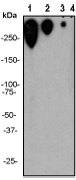
A. Immunoblot of different antigen-PEG preparations. Western Blot images using Anti-PEG 47 to detect BSA-PEG. Lane 1: 300 ng/mL; lane 2: 75 ng/mL; lane 3: 15 ng/mL; lane 4: 1.5 ng/mL.Application
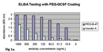
B. Comparison of anti-PEG-47 RabMAb and Vendor A mouse MAb in Direct ELISA assy. Goat anti-rabbit IgG-AP used for anti-PEG-47 detection; goat anti-mouse IgM-AP used for Vendor A MAb detection.Application
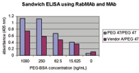
C. Comparison of sandwich ELISA using RabMAb/RabMAb (PEG 47/PEG 47) and MAb/RabMAb (Vendor A/PEG 47) for captureApplication
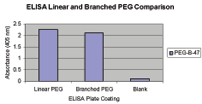
D. Comparison of 10 ug/well of activated linear (PEG5K) and branched (PEG40K) PEG using 5 ug/mL of anti-PEG-47 RabMAb in Direct ELISA assay.| Product Name | Polyethylene Glycol (PEG) Antibody |
|---|---|
| Antibody Type | Primary Antibodies |
| Clonality | Monoclonal |
|---|---|
| Clone Number | PEG-B-47 |
| Host Species | Rabbit |
| Tested Applications | WB |
| WB:1:500~1:2000 Notes:Optimal dilutions/concentrations should be determined by the researcher. |
|
| Species Reactivity | Human |
| Concentration | 1mg/ml |
Application
A. Immunoblot of different antigen-PEG preparations. Western Blot images using Anti-PEG 47 to detect BSA-PEG. Lane 1: 300 ng/mL; lane 2: 75 ng/mL; lane 3: 15 ng/mL; lane 4: 1.5 ng/mL.
Application
B. Comparison of anti-PEG-47 RabMAb and Vendor A mouse MAb in Direct ELISA assy. Goat anti-rabbit IgG-AP used for anti-PEG-47 detection; goat anti-mouse IgM-AP used for Vendor A MAb detection.
Application
C. Comparison of sandwich ELISA using RabMAb/RabMAb (PEG 47/PEG 47) and MAb/RabMAb (Vendor A/PEG 47) for capture
Application
D. Comparison of 10 ug/well of activated linear (PEG5K) and branched (PEG40K) PEG using 5 ug/mL of anti-PEG-47 RabMAb in Direct ELISA assay.| Application Notes | WB:1:500~1:2000 Notes:Optimal dilutions/concentrations should be determined by the researcher. |
|---|
| Form | Liquid |
|---|---|
| Storage Instructions | For short-term storage, store at 4° C. For long-term storage, aliquot and store at -20ºC or below. Avoid multiple freeze-thaw cycles. |
| Storage Buffer | 50 mM Tris-Glycine (pH 7.4), 0.15 M NaCl, 40% Glycerol, 0.01% sodium azide and 0.05% BSA. |


