Application
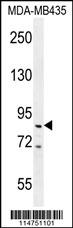
PPARGC Antibody western blot analysis in MDA-MB435 cell line lysates (35ug/lane).This demonstrates the PPARGC antibody detected the PPARGC protein (arrow).Application
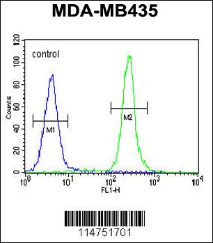
PPARGC Antibody flow cytometric analysis of MDA-MB435 cells (right histogram) compared to a negative control cell (left histogram).FITC-conjugated goat-anti-rabbit secondary antibodies were used for the analysis.| Product Name | PPARGC Antibody |
|---|---|
| Antibody Type | Primary Antibodies |
| Antigen Alias | PPARGC1A; LEM6; PGC1; PGC1A; PPARGC1; Peroxisome proliferator-activated receptor gamma coactivator 1-alpha; Ligand effect modulator 6 |
| Clonality | Polyclonal |
|---|---|
| Isotype | Ig |
| Host Species | Rabbit |
| Tested Applications | WBFC |
| WB:1:50~100 FC |
|
| Species Reactivity | Human |
| Concentration | 1mg/ml |
| Gene Synonyms | LEM6 PGC1 PGC1A PPARGC1 |
|---|---|
| Alternative Names | PPARGC1A LEM6 PGC1 PGC1A PPARGC1 Peroxisome proliferator-activated receptor gamma coactivator 1-alpha Ligand effect modulator 6 |
| Molecular Weight(MW) | 91027 Da |
| Function | Transcriptional coactivator for steroid receptors and nuclear receptors. Greatly increases the transcriptional activity of PPARG and thyroid hormone receptor on the uncoupling protein promoter. Can regulate key mitochondrial genes that contribute to the program of adaptive thermogenesis. Plays an essential role in metabolic reprogramming in response to dietary availability through coordination of the expression of a wide array of genes involved in glucose and fatty acid metabolism |
| Tissue Specificity | Heart, skeletal muscle, liver and kidney. Expressed at lower levels in brain and pancreas and at very low levels in the intestine and white adipose tissue. In skeletal muscle, levels were lower in obese than in lean subjects and fasting induced a 2-fold increase in levels in the skeletal muscle in obese subjects |
| Cellular Localization | Nucleus. |
| Entrez Gene | 10891 |
|---|---|
| Nucleotide Accession | NP_037393.1 |
Application
PPARGC Antibody western blot analysis in MDA-MB435 cell line lysates (35ug/lane).This demonstrates the PPARGC antibody detected the PPARGC protein (arrow).
Application
PPARGC Antibody flow cytometric analysis of MDA-MB435 cells (right histogram) compared to a negative control cell (left histogram).FITC-conjugated goat-anti-rabbit secondary antibodies were used for the analysis.| Application Notes | WB:1:50~100 FC |
|---|
| Form | Liquid |
|---|---|
| Storage Instructions | For short-term storage, store at 4° C. For long-term storage, aliquot and store at -20ºC or below. Avoid multiple freeze-thaw cycles. |
| Storage Buffer | Purified polyclonal antibody supplied in PBS with 0.09% (W/V) sodium azide. This antibody is prepared by Saturated Ammonium Sulfate (SAS) precipitation followed by dialysis against PBS. |


