Application
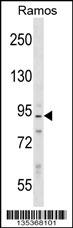
PPP1R9B Antibody (N-term) western blot analysis in Ramos cell line lysates (35ug/lane).This demonstrates the PPP1R9B antibody detected the PPP1R9B protein (arrow).| Product Name | PPP1R9B Antibody (N-term) |
|---|---|
| Antibody Type | Primary Antibodies |
| Antigen Alias | PPP1R9B; PPP1R6; Neurabin-2; Protein phosphatase 1 regulatory subunit 9B; Spinophilin; Neurabin-II |
| Clonality | Polyclonal |
|---|---|
| Isotype | Ig |
| Host Species | Rabbit |
| Tested Applications | WB |
| WB:1:100~500 |
|
| Species Reactivity | Human |
| Concentration | 1mg/ml |
| Gene Synonyms | PPP1R6 |
|---|---|
| Alternative Names | PPP1R9B PPP1R6 Neurabin-2 Protein phosphatase 1 regulatory subunit 9B Spinophilin Neurabin-II |
| Molecular Weight(MW) | 89192 Da |
| Function | Seems to act as a scaffold protein in multiple signaling pathways. Modulates excitatory synaptic transmission and dendritic spine morphology. Binds to actin filaments (F-actin) and shows cross-linking activity. Binds along the sides of the F-actin. May play an important role in linking the actin cytoskeleton to the plasma membrane at the synaptic junction. Believed to target protein phosphatase 1/PP1 to dendritic spines, which are rich in F-actin, and regulates its specificity toward ion channels and other substrates, such as AMPA-type and NMDA-type glutamate receptors. Plays a role in regulation of G-protein coupled receptor signaling, including dopamine D2 receptors and alpha- adrenergic receptors. May establish a signaling complex for dopaminergic neurotransmission through D2 receptors by linking receptors downstream signaling molecules and the actin cytoskeleton. Binds to ADRA1B and RGS2 and mediates regulation of ADRA1B signaling. May confer to Rac signaling specificity by binding to both, RacGEFs and Rac effector proteins. Probably regulates p70 S6 kinase activity by forming a complex with TIAM1 (By similarity). Required for hepatocyte growth factor (HGF)- induced cell migration |
| Tissue Specificity | This PPP1R9B antibody is generated from rabbits immunized with a KLH conjugated synthetic peptide between 23-53 amino acids from the N-terminal region of human PPP1R9B. |
| Cellular Localization | Cytoplasm, cytoskeleton (By similarity). Nucleus (By similarity). Cell projection, dendritic spine (By similarity). Cell junction, synapse. Cell junction, adherens junction (By similarity). Cytoplasm. Cell membrane. Cell projection, lamellipodium. Cell projection, filopodium. Cell projection, ruffle membrane. Note=Enriched at synapse and cadherin-based cell-cell adhesion sites. In neurons, both cytosolic and membrane-associated, and highly enriched in the postsynaptic density apposed to exitatory synapses. Colocalizes with PPP1R2 at actin-rich adherens junctions in epithelial cells and in dendritic spines (By similarity). Accumulates in the lamellipodium, filopodium and ruffle membrane in response to hepatocyte growth factor (HGF) treatment |
| Entrez Gene | 84687 |
|---|---|
| Nucleotide Accession | NP_115984.3 |
Application
PPP1R9B Antibody (N-term) western blot analysis in Ramos cell line lysates (35ug/lane).This demonstrates the PPP1R9B antibody detected the PPP1R9B protein (arrow).| Application Notes | WB:1:100~500 |
|---|
| Form | Liquid |
|---|---|
| Storage Instructions | For short-term storage, store at 4° C. For long-term storage, aliquot and store at -20ºC or below. Avoid multiple freeze-thaw cycles. |
| Storage Buffer | Purified polyclonal antibody supplied in PBS with 0.09% (W/V) sodium azide. This antibody is purified through a protein A column, followed by peptide affinity purification. |


