Application
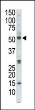
The anti-PRKAG2 Pab is used in Western blot to detect PRKAG2 in HL-60 cell lysate.Application
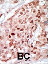
Formalin-fixed and paraffin-embedded human cancer tissue reacted with the primary antibody, which was peroxidase-conjugated to the secondary antibody, followed by DAB staining. This data demonstrates the use of this antibody for immunohistochemistry; clinical relevance has not been evaluated. BC = breast carcinoma; HC = hepatocarcinoma.Application
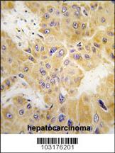
Formalin-fixed and paraffin-embedded human hepatocarcinoma tissue reacted with PRKAG2 Antibody (N-term) , which was peroxidase-conjugated to the secondary antibody, followed by DAB staining. This data demonstrates the use of this antibody for immunohistochemistry; clinical relevance has not been evaluated.Application
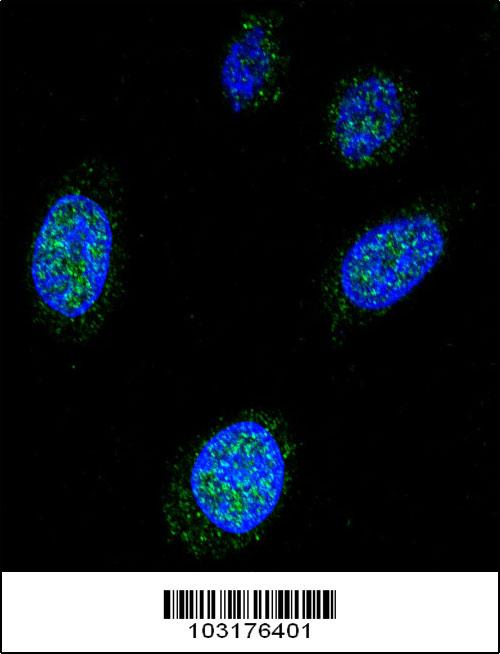
Confocal immunofluorescent analysis of PRKAG2 Antibody (N-term) (Cat#AP7049a) with Hela cell followed by Alexa Fluor 488-conjugated goat anti-rabbit lgG (green). DAPI was used to stain the cell nuclear (blue).| Product Name | PRKAG2 Antibody (N-term) |
|---|---|
| Antibody Type | Primary Antibodies |
| Antigen Alias | PRKAG2; 5'-AMP-activated protein kinase subunit gamma-2; H91620p |
| Clonality | Polyclonal |
|---|---|
| Isotype | Ig |
| Host Species | Rabbit |
| Tested Applications | WBIHCIF |
| WB:1:100~500 IHC |
|
| Species Reactivity | Human |
| Concentration | 1mg/ml |
| Alternative Names | PRKAG2 5'-AMP-activated protein kinase subunit gamma-2 H91620p |
|---|---|
| Molecular Weight(MW) | 63066 Da |
| Function | AMP/ATP-binding subunit of AMP-activated protein kinase (AMPK), an energy sensor protein kinase that plays a key role in regulating cellular energy metabolism. In response to reduction of intracellular ATP levels, AMPK activates energy-producing pathways and inhibits energy-consuming processes: inhibits protein, carbohydrate and lipid biosynthesis, as well as cell growth and proliferation. AMPK acts via direct phosphorylation of metabolic enzymes, and by longer-term effects via phosphorylation of transcription regulators. Also acts as a regulator of cellular polarity by remodeling the actin cytoskeleton; probably by indirectly activating myosin. Gamma non-catalytic subunit mediates binding to AMP, ADP and ATP, leading to activate or inhibit AMPK: AMP-binding results in allosteric activation of alpha catalytic subunit (PRKAA1 or PRKAA2) both by inducing phosphorylation and preventing dephosphorylation of catalytic subunits. ADP also stimulates phosphorylation, without stimulating already phosphorylated catalytic subunit. ATP promotes dephosphorylation of catalytic subunit, rendering the AMPK enzyme inactive |
| Tissue Specificity | Isoform B is ubiquitously expressed except in liver and thymus. The highest level is detected in heart with abundant expression in placenta and testis |
| Entrez Gene | 51422 |
|---|
Application
The anti-PRKAG2 Pab is used in Western blot to detect PRKAG2 in HL-60 cell lysate.
Application
Formalin-fixed and paraffin-embedded human cancer tissue reacted with the primary antibody, which was peroxidase-conjugated to the secondary antibody, followed by DAB staining. This data demonstrates the use of this antibody for immunohistochemistry; clinical relevance has not been evaluated. BC = breast carcinoma; HC = hepatocarcinoma.
Application
Formalin-fixed and paraffin-embedded human hepatocarcinoma tissue reacted with PRKAG2 Antibody (N-term) , which was peroxidase-conjugated to the secondary antibody, followed by DAB staining. This data demonstrates the use of this antibody for immunohistochemistry; clinical relevance has not been evaluated.
Application
Confocal immunofluorescent analysis of PRKAG2 Antibody (N-term) (Cat#AP7049a) with Hela cell followed by Alexa Fluor 488-conjugated goat anti-rabbit lgG (green). DAPI was used to stain the cell nuclear (blue).| Application Notes | WB:1:100~500 IHC |
|---|
| Form | Liquid |
|---|---|
| Storage Instructions | For short-term storage, store at 4° C. For long-term storage, aliquot and store at -20ºC or below. Avoid multiple freeze-thaw cycles. |
| Storage Buffer | Purified polyclonal antibody supplied in PBS with 0.09% (W/V) sodium azide. This antibody is purified through a protein G column, eluted with high and low pH buffers and neutralized immediately, followed by dialysis against PBS. |


