Application
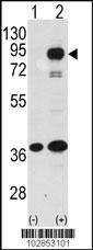
Western blot analysis of PROX1 (arrow) using rabbit polyclonal PROX1 Antibody .293 cell lysates (2 ug/lane) either nontransfected (Lane 1) or transiently transfected with the PROX1 gene (Lane 2) (Origene Technologies).Application
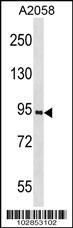
PROX1 Antibody (K507) western blot analysis in A2058 cell line lysates (35ug/lane).This demonstrates the PROX1 antibody detected the PROX1 protein (arrow).Application
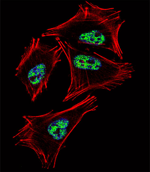
Fluorescent confocal image of A2058 cell stained with PROX1 Antibody (C-term)(Cat#AP2035b).A2058 cells were fixed with 4% PFA (20 min), permeabilized with Triton X-100 (0.1%, 10 min), then incubated with PROX1 primary antibody (1:25, 1 h at 37℃). For secondary antibody, Alexa Fluor® 488 conjugated donkey anti-rabbit antibody (green) was used (1:400, 50 min at 37℃).Cytoplasmic actin was counterstained with Alexa Fluor® 555 (red) conjugated Phalloidin (7units/ml, 1 h at 37℃). Nuclei were counterstained with DAPI (blue) (10 µg/ml, 10 min). PROX1 immunoreactivity is localized to Nucleus significantly.| Product Name | PROX1 Antibody (C-term) |
|---|---|
| Antibody Type | Primary Antibodies |
| Antigen Alias | PROX1; Prospero homeobox protein 1; Homeobox prospero-like protein PROX1 |
| Clonality | Polyclonal |
|---|---|
| Isotype | Ig |
| Host Species | Rabbit |
| Tested Applications | WBIF |
| WB:1:50~100 IF |
|
| Species Reactivity | Human |
| Concentration | 1mg/ml |
| Alternative Names | PROX1 Prospero homeobox protein 1 Homeobox prospero-like protein PROX1 |
|---|---|
| Molecular Weight(MW) | 83203 Da |
| Function | May play a fundamental role in early development of CNS. May regulate gene expression and development of postmitotic undifferentiated young neurons (By similarity) |
| Tissue Specificity | Most actively expressed in the developing lens. Detected also in embryonic brain, lung, liver and kidney. In adult, it is more abundant in heart and liver than in brain, skeletal muscle, kidney and pancreas |
| Cellular Localization | Nucleus (Probable). |
Application
Western blot analysis of PROX1 (arrow) using rabbit polyclonal PROX1 Antibody .293 cell lysates (2 ug/lane) either nontransfected (Lane 1) or transiently transfected with the PROX1 gene (Lane 2) (Origene Technologies).
Application
PROX1 Antibody (K507) western blot analysis in A2058 cell line lysates (35ug/lane).This demonstrates the PROX1 antibody detected the PROX1 protein (arrow).
Application
Fluorescent confocal image of A2058 cell stained with PROX1 Antibody (C-term)(Cat#AP2035b).A2058 cells were fixed with 4% PFA (20 min), permeabilized with Triton X-100 (0.1%, 10 min), then incubated with PROX1 primary antibody (1:25, 1 h at 37℃). For secondary antibody, Alexa Fluor® 488 conjugated donkey anti-rabbit antibody (green) was used (1:400, 50 min at 37℃).Cytoplasmic actin was counterstained with Alexa Fluor® 555 (red) conjugated Phalloidin (7units/ml, 1 h at 37℃). Nuclei were counterstained with DAPI (blue) (10 µg/ml, 10 min). PROX1 immunoreactivity is localized to Nucleus significantly.| Application Notes | WB:1:50~100 IF |
|---|
| Form | Liquid |
|---|---|
| Storage Instructions | For short-term storage, store at 4° C. For long-term storage, aliquot and store at -20ºC or below. Avoid multiple freeze-thaw cycles. |
| Storage Buffer | Purified polyclonal antibody supplied in PBS with 0.09% (W/V) sodium azide. This antibody is purified through a protein G column, eluted with high and low pH buffers and neutralized immediately, followed by dialysis against PBS. |


