Application
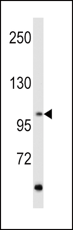
Western blot analysis of hRET-G28 in MCF7 cell line lysates (35ug/lane). RET (arrow) was detected using the purified Pab.Application
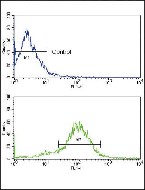
Flow cytometric analysis of MCF-7 cells using RET Antibody (N-term Q28) (bottom histogram) compared to a negative control cell (top histogram). FITC-conjugated goat-anti-rabbit secondary antibodies were used for the analysis.| Product Name | RET Antibody (N-term Q28) |
|---|---|
| Antibody Type | Primary Antibodies |
| Antigen Alias | RET; CDHF12; CDHR16; PTC; RET51; Proto-oncogene tyrosine-protein kinase receptor Ret; Cadherin family member 12; Proto-oncogene c-Ret; Soluble RET kinase fragment; Extracellular cell-membrane anchored RET cadherin 120 kDa fragment |
| Clonality | Polyclonal |
|---|---|
| Isotype | Ig |
| Host Species | Rabbit |
| Tested Applications | WBFC |
| WB:1:100~500 FC |
|
| Species Reactivity | Human |
| Concentration | 1mg/ml |
| Gene Synonyms | CDHF12 CDHR16 PTC RET51 |
|---|---|
| Alternative Names | RET CDHF12 CDHR16 PTC RET51 Proto-oncogene tyrosine-protein kinase receptor Ret Cadherin family member 12 Proto-oncogene c-Ret Soluble RET kinase fragment Extracellular cell-membrane anchored RET cadherin 120 kDa fragment |
| Molecular Weight(MW) | 124319 Da |
| Function | Receptor tyrosine-protein kinase involved in numerous cellular mechanisms including cell proliferation, neuronal navigation, cell migration, and cell differentiation upon binding with glial cell derived neurotrophic factor family ligands Phosphorylates PTK2/FAK1. Regulates both cell death/survival balance and positional information. Required for the molecular mechanisms orchestration during intestine organogenesis; involved in the development of enteric nervous system and renal organogenesis during embryonic life, and promotes the formation of Peyer's patch-like structures, a major component of the gut- associated lymphoid tissue. Modulates cell adhesion via its cleavage by caspase in sympathetic neurons and mediates cell migration in an integrin (e.g. ITGB1 and ITGB3)-dependent manner Involved in the development of the neural crest. Active in the absence of ligand, triggering apoptosis through a mechanism that requires receptor intracellular caspase cleavage. Acts as a dependence receptor; in the presence of the ligand GDNF in somatotrophs (within pituitary), promotes survival and down regulates growth hormone (GH) production, but triggers apoptosis in absence of GDNF. Regulates nociceptor survival and size Triggers the differentiation of rapidly adapting (RA) mechanoreceptors. Mediator of several diseases such as neuroendocrine cancers; these diseases are characterized by aberrant integrins-regulated cell migration |
| Tissue Specificity | This RET antibody is generated from rabbits immunized with a KLH conjugated synthetic peptide between 21~51 amino acids from the N-terminal region of human RET. |
| Cellular Localization | Cell membrane; Single-pass type I membrane protein. Endosome membrane; Single-pass type I membrane protein |
| Entrez Gene | 5979 |
|---|
Application
Western blot analysis of hRET-G28 in MCF7 cell line lysates (35ug/lane). RET (arrow) was detected using the purified Pab.
Application
Flow cytometric analysis of MCF-7 cells using RET Antibody (N-term Q28) (bottom histogram) compared to a negative control cell (top histogram). FITC-conjugated goat-anti-rabbit secondary antibodies were used for the analysis.| Application Notes | WB:1:100~500 FC |
|---|
| Form | Liquid |
|---|---|
| Storage Instructions | For short-term storage, store at 4° C. For long-term storage, aliquot and store at -20ºC or below. Avoid multiple freeze-thaw cycles. |
| Storage Buffer | Purified polyclonal antibody supplied in PBS with 0.09% (W/V) sodium azide. This antibody is purified through a protein G column, eluted with high and low pH buffers and neutralized immediately, followed by dialysis against PBS. |


