Application
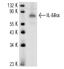
Western blot analysis of SAP Antibody (Center E300) (Cat. 3AP7398c) in Y79,A2058,CEM cell line lysates(35ug/lane). SAP (arrow) was detected using the purified Pab.| Product Name | SAP Antibody (Center E300) |
|---|---|
| Antibody Type | Primary Antibodies |
| Antigen Alias | PSAP; GLBA; SAP1; Proactivator polypeptide; Saposin-A; Protein A; Saposin-B-Val; Saposin-B; Cerebroside sulfate activator; Dispersin; Sphingolipid activator protein 1; Sulfatide/GM1 activator; Saposin-C; A1 activator; Co-beta-glucosidase; Glucosylceramidas |
| Clonality | Polyclonal |
|---|---|
| Isotype | Ig |
| Host Species | Rabbit |
| Tested Applications | WB |
| WB:1:50~100 |
|
| Species Reactivity | Human |
| Concentration | 1mg/ml |
| Gene Synonyms | GLBA SAP1 |
|---|---|
| Alternative Names | PSAP GLBA SAP1 Proactivator polypeptide Saposin-A Protein A Saposin-B-Val Saposin-B Cerebroside sulfate activator Dispersin Sphingolipid activator protein 1 Sulfatide/GM1 activator Saposin-C A1 activator Co-beta-glucosidase Glucosylceramidas |
| Molecular Weight(MW) | 58113 Da |
| Function | The lysosomal degradation of sphingolipids takes place by the sequential action of specific hydrolases. Some of these enzymes require specific low-molecular mass, non-enzymic proteins: the sphingolipids activator proteins (coproteins) Saposin-B stimulates the hydrolysis of galacto- cerebroside sulfate by arylsulfatase A (EC 3.1.6.8), GM1 gangliosides by beta-galactosidase (EC 3.2.1.23) and globotriaosylceramide by alpha-galactosidase A (EC 3.2.1.22) Saposin-B forms a solubilizing complex with the substrates of the sphingolipid hydrolases |
| Tissue Specificity | This SAP antibody is generated from rabbits immunized with a KLH conjugated synthetic peptide between 293~322 amino acids from the Center region of human SAP. |
| Cellular Localization | Lysosome. |
| Entrez Gene | 5660 |
|---|
Application
Western blot analysis of SAP Antibody (Center E300) (Cat. 3AP7398c) in Y79,A2058,CEM cell line lysates(35ug/lane). SAP (arrow) was detected using the purified Pab.| Application Notes | WB:1:50~100 |
|---|
| Form | Liquid |
|---|---|
| Storage Instructions | For short-term storage, store at 4° C. For long-term storage, aliquot and store at -20ºC or below. Avoid multiple freeze-thaw cycles. |
| Storage Buffer | Purified polyclonal antibody supplied in PBS with 0.09% (W/V) sodium azide. This antibody is prepared by Saturated Ammonium Sulfate (SAS) precipitation followed by dialysis against PBS. |


