Application
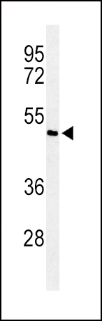
Western blot analysis of Smad7 Antibody (Center) in ZR-75-1 cell line lysates (35ug/lane). Smad7 (arrow) was detected using the purified Pab.Application
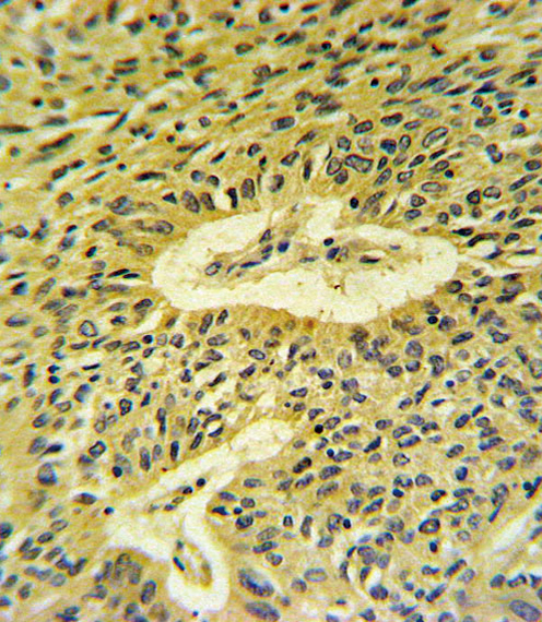
Smad7 Antibody (Center) IHC analysis in formalin fixed and paraffin embedded lung carcinoma followed by peroxidase conjugation of the secondary antibody and DAB staining. This data demonstrates the use of the Smad7 Antibody (Center) for immunohistochemistry. Clinical relevance has not been evaluated.Application
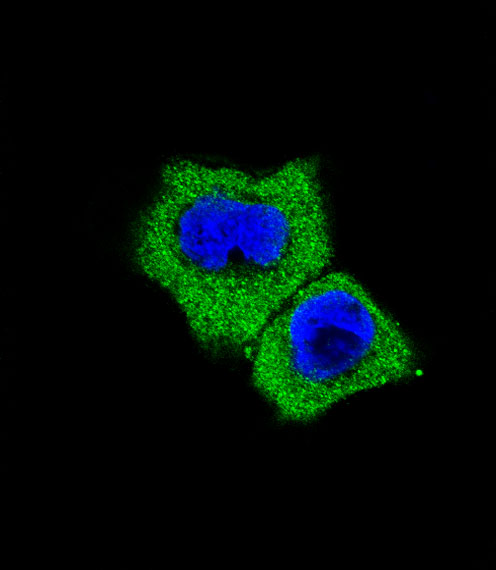
Fluorescent confocal image of A431 cell stained with Smad7 Antibody (Center)(Cat#AP6753c). A431 cells were fixed with 4% PFA (20 min), permeabilized with Triton X-100 (0.1%, 10 min), then incubated with Smad7 primary antibody (1:25, 1 h at 37℃). For secondary antibody, Alexa Fluor® 488 conjugated donkey anti-rabbit antibody (green) was used (1:400, 50 min at 37℃).Cytoplasmic actin was counterstained with Alexa Fluor® 555 (red) conjugated Phalloidin (7units/ml, 1 h at 37℃). Nuclei were counterstained with DAPI (blue) (10 µg/ml, 10 min).Smad7 immunoreactivity is localized to cytoplasm significantly.| Product Name | Smad7 Antibody (Center) |
|---|---|
| Antibody Type | Primary Antibodies |
| Antigen Alias | SMAD7; MADH7; MADH8; Mothers against decapentaplegic homolog 7; Mothers against decapentaplegic homolog 8; SMAD family member 7 |
| Clonality | Polyclonal |
|---|---|
| Isotype | Ig |
| Host Species | Rabbit |
| Tested Applications | WBIHCIF |
| WB:1:100~500 IHC |
|
| Species Reactivity | Human |
| Concentration | 1mg/ml |
| Gene Synonyms | MADH7 MADH8 |
|---|---|
| Alternative Names | SMAD7 MADH7 MADH8 Mothers against decapentaplegic homolog 7 Mothers against decapentaplegic homolog 8 SMAD family member 7 |
| Molecular Weight(MW) | 46426 Da |
| Function | Antagonist of signaling by TGF-beta (transforming growth factor) type 1 receptor superfamily members; has been shown to inhibit TGF-beta (Transforming growth factor) and activin signaling by associating with their receptors thus preventing SMAD2 access. Functions as an adapter to recruit SMURF2 to the TGF-beta receptor complex. Also acts by recruiting the PPP1R15A- PP1 complex to TGFBR1, which promotes its dephosphorylation Positively regulates PDPK1 kinase activity by stimulating its dissociation from the 14-3-3 protein YWHAQ which acts as a negative regulator (By similarity) |
| Tissue Specificity | Ubiquitous with higher expression in the lung and vascular endothelium |
| Cellular Localization | Nucleus. Cytoplasm. Note=Interaction with NEDD4L or RNF111 or induces translocation from the nucleus to the cytoplasm. TGF-beta stimulates its translocation from the nucleus to the cytoplasm. PDPK1 inhibits its translocation from the nucleus to the cytoplasm in response to TGF-beta |
| Entrez Gene | 4092 |
|---|
Application
Western blot analysis of Smad7 Antibody (Center) in ZR-75-1 cell line lysates (35ug/lane). Smad7 (arrow) was detected using the purified Pab.
Application
Smad7 Antibody (Center) IHC analysis in formalin fixed and paraffin embedded lung carcinoma followed by peroxidase conjugation of the secondary antibody and DAB staining. This data demonstrates the use of the Smad7 Antibody (Center) for immunohistochemistry. Clinical relevance has not been evaluated.
Application
Fluorescent confocal image of A431 cell stained with Smad7 Antibody (Center)(Cat#AP6753c). A431 cells were fixed with 4% PFA (20 min), permeabilized with Triton X-100 (0.1%, 10 min), then incubated with Smad7 primary antibody (1:25, 1 h at 37℃). For secondary antibody, Alexa Fluor® 488 conjugated donkey anti-rabbit antibody (green) was used (1:400, 50 min at 37℃).Cytoplasmic actin was counterstained with Alexa Fluor® 555 (red) conjugated Phalloidin (7units/ml, 1 h at 37℃). Nuclei were counterstained with DAPI (blue) (10 µg/ml, 10 min).Smad7 immunoreactivity is localized to cytoplasm significantly.| Application Notes | WB:1:100~500 IHC |
|---|
| Form | Liquid |
|---|---|
| Storage Instructions | For short-term storage, store at 4° C. For long-term storage, aliquot and store at -20ºC or below. Avoid multiple freeze-thaw cycles. |
| Storage Buffer | Purified polyclonal antibody supplied in PBS with 0.09% (W/V) sodium azide. This antibody is prepared by Saturated Ammonium Sulfate (SAS) precipitation followed by dialysis against PBS. |


