Application
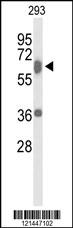
Western blot analysis of TAB1 Antibody (Center) in 293 cell line lysates (35ug/lane). TAB1 (arrow) was detected using the purified Pab.Application
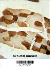
TAB1 Antibody (Center) immunohistochemistry analysis in formalin fixed and paraffin embedded human skeletal muscle followed by peroxidase conjugation of the secondary antibody and DAB staining. This data demonstrates the use of the TAB1 Antibody (Center) for immunohistochemistry. Clinical relevance has not been evaluated.Application
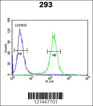
TAB1 Antibody (Center) flow cytometric analysis of 293 cells (right histogram) compared to a negative control cell (left histogram).FITC-conjugated goat-anti-rabbit secondary antibodies were used for the analysis.| Product Name | TAB1 Antibody (Center) |
|---|---|
| Antibody Type | Primary Antibodies |
| Antigen Alias | TAB1; MAP3K7IP1; TGF-beta-activated kinase 1 and MAP3K7-binding protein 1; Mitogen-activated protein kinase kinase kinase 7-interacting protein 1; TGF-beta-activated kinase 1-binding protein 1 |
| Clonality | Polyclonal |
|---|---|
| Isotype | Ig |
| Host Species | Rabbit |
| Tested Applications | WBIHCFC |
| WB:1:100~500 IHC |
|
| Species Reactivity | Human |
| Concentration | 1mg/ml |
| Gene Synonyms | MAP3K7IP1 |
|---|---|
| Alternative Names | TAB1 MAP3K7IP1 TGF-beta-activated kinase 1 and MAP3K7-binding protein 1 Mitogen-activated protein kinase kinase kinase 7-interacting protein 1 TGF-beta-activated kinase 1-binding protein 1 |
| Molecular Weight(MW) | 54644 Da |
| Function | May be an important signaling intermediate between TGFB receptors and MAP3K7/TAK1. May play an important role in mammalian embryogenesis |
| Tissue Specificity | Ubiquitous. |
| Entrez Gene | 10454 |
|---|
Application
Western blot analysis of TAB1 Antibody (Center) in 293 cell line lysates (35ug/lane). TAB1 (arrow) was detected using the purified Pab.
Application
TAB1 Antibody (Center) immunohistochemistry analysis in formalin fixed and paraffin embedded human skeletal muscle followed by peroxidase conjugation of the secondary antibody and DAB staining. This data demonstrates the use of the TAB1 Antibody (Center) for immunohistochemistry. Clinical relevance has not been evaluated.
Application
TAB1 Antibody (Center) flow cytometric analysis of 293 cells (right histogram) compared to a negative control cell (left histogram).FITC-conjugated goat-anti-rabbit secondary antibodies were used for the analysis.| Application Notes | WB:1:100~500 IHC |
|---|
| Form | Liquid |
|---|---|
| Storage Instructions | For short-term storage, store at 4° C. For long-term storage, aliquot and store at -20ºC or below. Avoid multiple freeze-thaw cycles. |
| Storage Buffer | Purified polyclonal antibody supplied in PBS with 0.09% (W/V) sodium azide. This antibody is purified through a protein A column, followed by peptide affinity purification. |


 Data sheet for OM222473